- Clinical Technology
- Adult Immunization
- Hepatology
- Pediatric Immunization
- Screening
- Psychiatry
- Allergy
- Women's Health
- Cardiology
- Pediatrics
- Dermatology
- Endocrinology
- Pain Management
- Gastroenterology
- Infectious Disease
- Obesity Medicine
- Rheumatology
- Nephrology
- Neurology
- Pulmonology
Respiratory Disorders-A Photo Essay
Acute bronchiolitis, COPD, giant bullous emphysema, hypersensitivity pneumonitis, and pulmonary tuberculosis may present diagnostic challenges.
This chest radiograph shows a normal heart contour with an upper border merging with thymic tissue, a right middle lobe infiltrate, and peribronchial cuffing in both lung fields. The infant's intermittent wheezing, stable cardiac lesions, and acute pulmonary infiltrate favor a diagnosis of recurrent respiratory illness, manifested as acute bronchiolitis and right middle lobe pneumonia.
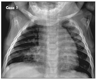
Image courtesy Linh Thi My Ha, MD and Golder N. Wilson, MD, PhD.
Click here for the next image
Parametric response mapping, used to analyze lung CT scans, helps physicians distinguish between early-stage airway damage and emphysema. In lung images of a healthy person, 2 persons with mild to moderate chronic obstructive pulmonary disease, and one with severe emphysema (left to right), green is healthy tissue, yellow is small airway damage, and red is more severe damage.

Image courtesy of the Center for Molecular Imaging, University of Michigan.
Click here for the next image
A CT scan of the thorax was ordered to help determine whether a recurrent pulmonary embolism may have contributed to a 54-year-old man's arrhythmia. He also had a history of giant bullous emphysema, or “vanishing lung syndrome,” a rare condition caused by paraseptal emphysematous bullae that coalesce and eventually compress the lung parenchyma. The bullae can be mistaken for a pneumothorax on a standard chest film.
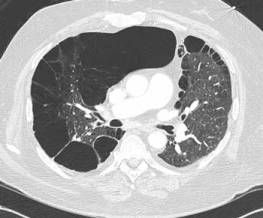
Image courtesy of Mark Masciocchi, MD, Shashank Jain, MD, and Anthony Donato, MD.
Click here for the next image
Drug-induced hypersensitivity pneumonitis is a clinically distinct pulmonary syndrome characterized by a complex immunological reaction. This lung biopsy specimen shows loosely formed granulomas near the terminal bronchioles and lymphocytic and plasma cell infiltration of the alveolar walls. Chronic interstitial pneumonia, loose granulomas, and areas of organizing pneumonia are consistent with hypersensitivity pneumonitis.
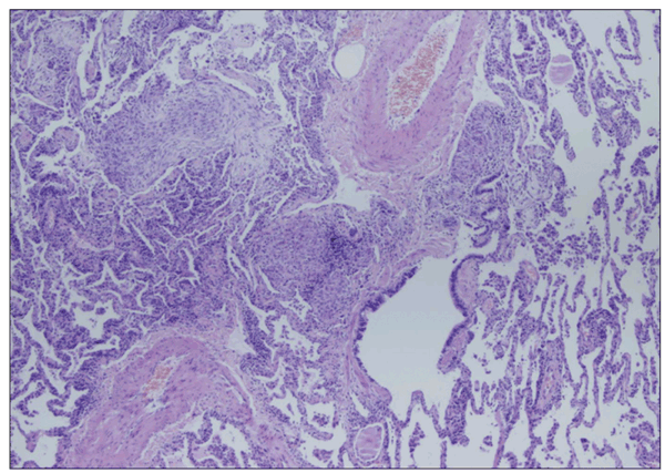
Image courtesy of Krish Bhadra, MD and Benjamin T. Suratt, MD
Click here for the next image
A chest radiograph from a baby girl with a history of difficulty in breathing showed a hyperinflated right lung with a shift of the mediastinal contents into the left hemithorax (A). A chest CT scan showed diffuse mediastinal and right hilar adenopathy (B). The case emphasizes the difficulty in diagnosing pulmonary tuberculosis in an infant with a nonspecific presentation and no travel or contact history.
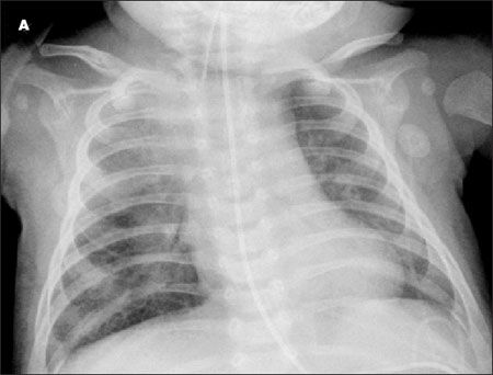
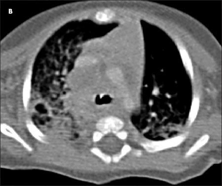
Image courtesy of Mansi B. Mehta, MD, Christopher Young, MD, and Sharda Udassi, MD.
Click here for the next image
An open lung biopsy performed 5 days after a previously healthy young man with hemoptysis was intubated showed bronchiolitis obliterans organizing pneumonia, characterized by granulation tissue in the bronchiolar lumen, alveolar ducts, and some alveoli. Chest radiographs typically show bilateral patchy alveolar infiltrates and nodules. Lung biopsy via video-assisted thoracoscopic surgery is preferred for diagnosis.
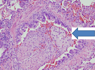
Image courtesy of Zynab Hassan, MD, Isaac Goldberg, MD, and Clare Hawkins, MD.
Click here to return to the first image.
