- Clinical Technology
- Adult Immunization
- Hepatology
- Pediatric Immunization
- Screening
- Psychiatry
- Allergy
- Women's Health
- Cardiology
- Pediatrics
- Dermatology
- Endocrinology
- Pain Management
- Gastroenterology
- Infectious Disease
- Obesity Medicine
- Rheumatology
- Nephrology
- Neurology
- Pulmonology
Refractory Ulcer:What to Do Next?
ABSTRACT: Undiagnosed or persistent Helicobacter pylori infection and surreptitious or unrecognized NSAID use are the most common causes of refractory peptic ulcers. The use of antibiotics, bismuth, or proton pump inhibitors (PPIs) suppresses the H pylori bacterial load and may obscure the diagnosis. H pylori infections have also become more difficult to cure because of increased antibiotic resistance. For refractory infection, select an antibiotic based on in vitro susceptibility testing. When this is not available, combination therapy with a PPI, tetracycline, metronidazole, and bismuth is often effective. To detect surreptitious or inadvertent NSAID use, review the drug history in detail. When there is any doubt about such use, check platelet cyclooxygenase function.
ABSTRACT: Undiagnosed or persistent Helicobacter pylori infection and surreptitious or unrecognized NSAID use are the most common causes of refractory peptic ulcers. The use of antibiotics, bismuth, or proton pump inhibitors (PPIs) suppresses the H pylori bacterial load and may obscure the diagnosis. H pylori infections have also become more difficult to cure because of increased antibiotic resistance. For refractory infection, select an antibiotic based on in vitro susceptibility testing. When this is not available, combination therapy with a PPI, tetracycline, metronidazole, and bismuth is often effective. To detect surreptitious or inadvertent NSAID use, review the drug history in detail. When there is any doubt about such use, check platelet cyclooxygenase function.
The development of effective antimicrobial therapy to eradicate Helicobacter pylori infection and the widespread use of potent proton pump inhibitors (PPIs) have revolutionized the management of peptic ulcer disease. Most peptic ulcers heal within 1 to 2 months of conventional anti- secretory treatment; however, 5% to 10% fail to heal within this period or persist despite prolonged antisecretory drug therapy.1 The small number of ulcers that persist despite conventional treatment are considered refractory.2,3
Here we review the causes of refractory ulcer and describe specific management strategies.
PEPTIC ULCER DISEASE: A BRIEF OVERVIEW
Peptic ulcerations are excavated mucosal defects that result from destruction of epithelial cells by the caustic effects of acid and pepsin in the GI lumen. Ulcers have been defined histologically as necrotic mucosal defects that extend through the muscularis mucosae and into the submucosa or deeper layers (Figure). Necrotic defects that are more superficial are considered erosions.4,5 Ulcers can also be characterized as acute or chronic depending on the absence or presence of fibrosis. This article focuses on chronic peptic ulcers.


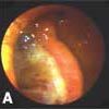
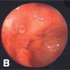
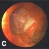
Figure – Endoscopy revealed a large, deep, slow-healing, NSAID-associated ulcer on the anterior wall of the antrum (A). The paient's test results for Helicobacter pylori were negative. Six months later, after continuous twice-a-day omeprazole therapy, the ulcer had almost completely healed (B). Within a week of restarting low-dose NSAID therapy (the proton pump inhibitor was discontinued), the ulcer recurred (C). Histological findings after resection were typical of gastric ulcer.
The most common causes of peptic ulcer disease are H pylori infection and NSAID use (Table).6 Clinically, the natural history of peptic ulcer disease is one of exacerbation and remission. Unless the causative factor is eliminated, the recurrence rate ranges between 60% and 100% per year. Elimination of the causative factor is expected to cure the disease and prevent recurrence.
Table – Causes and associations of peptic ulcer disease
DEFINITION OF REFRACTORY ULCER
Lanas and colleagues7 defined refractory ulcers based on the type and duration of treatment. Refractory duodenal ulcers were those that did not heal after 8 weeks of full-dose H2-receptor antagonist therapy or after 6 weeks of PPI therapy. Refractory gastric ulcers were defined as those that did not heal after 12 weeks of full-dose H2-receptor antagonist therapy or 8 weeks of PPI therapy. In this article, we define refractory ulcers as those that do not heal after 12 weeks of therapy with PPIs or those that recur rapidly after antisecretory drug therapy is discontinued.
ULCER HEALING
Factors that facilitate healing. Ulcer healing is initiated by the secretion of growth factors in the ulcer margin and ulcer bed; the mucosal defect is filled with cells that migrate from the ulcer margin and with connective tissue.8,9 The epidermal growth factor, transforming growth factor-α, hepatocyte growth factor, and trefoil factors have been implicated in this process. These growth factors regulate cell proliferation, migration, differentiation, and secretion and degradation of the extracellular matrix-all of which are essential for tissue healing.9
Gastric mucosal blood flow is also important for ulcer healing. In one study, the refractory ulcers persistently had significantly lower mucosal blood flow than that of normal stomachs and healing ulcers.10
Factors that impede healing. Clinical markers associated with slow ulcer healing include11,12:
• Large ulcer size.
• Persistent ulcer symptoms.
• Alcohol use.
• Cigarette smoking.
• NSAID use.
In one study, 90% of aspirin-associated gastric ulcers of less than 0.5 cm in diameter healed within 8 weeks of treatment with cimetidine and antacids despite the continued use of aspirin, whereas only 25% of gastric ulcers of greater than 0.5 cm in diameter healed in this same setting.13
NSAID use is the most common factor associated with slow healing and rapid recurrence of peptic ulcers. NSAIDs are thought to delay healing by interfering with the action of growth factors, decreasing epithelial cell proliferation in the ulcer margin, decreasing angiogenesis in the ulcer bed, and slowing maturation of the granulation tissue.8
CAUSES OF REFRACTORY ULCER
Among the causes of refractory or frequently recurring ulcers are persistent H pylori infection14; use of NSAIDs or other ulcerogenic drugs; pathological gastric acid hypersecretion; and other infectious diseases, such as herpes simplex, Cytomegalovirus infection, syphilis, and tuberculosis. Included in the differential diagnosis are diseases that may mimic peptic ulcers, such as Crohn disease and various tumors.3,15-19 The most common causes of refractory ulcer are undiagnosed and persistent H pylori infection and surreptitious or inadvertent NSAID use.17
H pylori infection. The use of antibiotics, bismuth, or PPIs suppresses the H pylori bacterial load and may obscure the diagnosis. Use of these agents is associated with false-negative results for tests of active infection, including rapid urease tests, culture, histology, urea breath tests, and stool antigen tests.
PPIs are associated both with a marked reduction in the H pylori bacterial load, especially in the antrum, and with normalization of antral histological findings. Thus, to detect infection, biopsy of the gastric corpus mucosa as well as the antrum should be performed. Because the results of serological tests are not affected by drugs that reduce the bacterial load, testing for H pylori antibodies is recommended when the diagnosis is in doubt. Consider switching the patient from a PPI to an H2-receptor antagonist several weeks before either noninvasive testing (stool antigen or urea breath tests) or endoscopy for histology and possibly culture; H2 blockers do not affect H pylori status.
H pylori infections have become more difficult to cure in part because of the increased prevalence of antibiotic resistance. Consider local resis- tance patterns when treating these infections empirically.20 For refractory H pylori infection, select an antibiotic based on in vitro susceptibility testing. When this is not available, combination therapy with a PPI, tetracycline, metronidazole, and bismuth is often effective. A number of references list different options for salvage therapies.6,15,21
NSAID use. NSAIDs (including aspirin) are an important cause of peptic ulcers.22 Although NSAIDs are more likely to produce gastric ulcers than duodenal ulcers, they are a prominent cause of giant and refractory duodenal ulcers.
The ratio of H pylori–associated ulcers to NSAID-associated ulcers depends on local prevalence of H pylori infection and NSAID use. In populations with a low H pylori infection rate and a high frequency of NSAID use (eg, in elderly persons), NSAIDs have become the major risk factor for the development of peptic ulcer. This is of particular concern in elderly persons because they are less able to tolerate ulcer-related complications, such as hemorrhage and perforation.23
Inhibition of the isoenzyme cyclooxygenase (COX)-1 is largely responsible for NSAID-associated ulcers. It was thought that highly selective COX-2 inhibitors would help reduce the problem; however, their use has been limited by their potential to increase cardiovascular events and by their reduced or eliminated selectivity with concomitant aspirin use.
Surreptitious use of aspirin or NSAIDs can be detected by testing for platelet COX activity.3 This test has been shown to be a potent and sensitive method to detect current aspirin use.24 In one prospective study, current NSAID use (of which 89% was aspirin) was detected in 80% of patients with upper or lower GI bleeding; 21.5% more aspirin users were detected by the COX test than by history alone.24
NSAID use can be inadvertent. Because many over-the-counter medications (eg, cough-cold remedies) contain NSAIDs, inquire about the use of these and other products from health food or vitamin stores during the history taking. Although the full range of ulcerogenic drugs is unknown, consider solid-dose forms of potassium and bisphosphonates, such as alendronate and risedronate, as potential culprits.25
Zollinger-Ellison syndrome (ZES). This syndrome may be a hidden cause of refractory peptic ulcers. ZES is characterized by peptic ulcers of the upper GI tract, which are difficult to healand rapidly recur when antisecretory drug therapy is withdrawn. Diarrhea also may be associated with the marked gastric acid hypersecretion and often resolves with PPI therapy and recurs when antisecretory drugs are withdrawn. H pylori infection is typically absent.
Patients with ZES classically have very healthy–appearing gastric mucosa. Ulcers are often present in the duodenum; an ulcer in the postbulbar region or beyond is highly suggestive of ZES. Gastric ulcers are rare; when ulcers are present in the body of the stomach, the diagnosis of ZES is unlikely. Erosive gastroesophageal reflux is common.
Diagnosis. ZES is caused by gastrin-secreting tumors (gastrinoma).26 A fasting plasma gastrin level greater than 1000 pg/mL or a basal acid output greater than 15 mEq/h suggests the diagnosis. In the United States, the estimated frequency of ZES is 0.1 to 3 per million persons. The percentage of persons with refractory ulcer who have ZES is unknown.
When ZES is suspected, a fasting serum gastrin level is measured theoretically when antisecretory medications are discontinued. Sometimes, it is difficult to convince the patient to stop PPI therapy. If the gastrin level is significantly elevated (eg, greater than 400 pg/mL), measure gastric acidity, because hypochlorhydria causes feedback stimulation of antral gastrin secretion.
In suspected cases of ZES with mild hypergastrinemia, the secretin stimulation test may be useful.15 The diagnosis of ZES may be difficult in the setting of potent medical acid suppression therapy, especially in the presence of atrophic gastritis.15 In our experience, the conditions can be differentiated clinically. For instance, ZES alone is associated with duodenal ulcers, thick gastric folds, enterochromaffin cell hyperplasia, thick and healthy-appearing gastric mucosal histology with prominent parietal cells, and a low gastric pH. In contrast, PPI-associated hypergastrinemia is associated with an atrophic-appearing gastric corpus, thick secretions, hypochlorhydria or achlorhydria, few parietal cells, and gastric ulcers.
In patients with ZES, it is important to determine whether the condition is part of the multiple endocrine neoplasia syndrome, in which the tumors are typically multifocal and malignant. If not, localization of the tumor or metastases is essential. Somatostatin-receptor scintigraphy can localize the tumor in 80% of cases and can identify tumors located in sites other than the pancreas or duodenum.27 Endoscopic ultrasonography is another useful method; it has a sensitivity as high as 79% to 93% and a specificity as high as 93%.
Treatment. The 2 principal therapeutic strategies are to control the gastric acid hypersecretion and to inhibit neoplasia.28 Surgical excision of the gastrinoma is the best treatment. PPIs are the drugs of choice for patients with ZES and are administered at sufficiently high dosages to reduce the basal acid secretion to less than 10 mEq/hfor the hour before the next dose.26
Idiopathic gastric acid hypersecretion. Refractory ulcers with no associated risk factors are rare but not exceptional. Smoking and genetic background seem to be important factors in patients with idiopathic refractory ulcers.22 Idiopathic gastric acid hypersecretion is associated with duodenal ulcers and with the absence of H pylori infection. The condition may be an important consideration in a few patients in whom ZES can be excluded. To rule out ZES, Lewis29 defined idiopathic gastric acid hypersecretion as a basal acid output of more than 10 mEq/h in the absence of an elevated fasting serum gastrin level (or a negative secretin test, if the gastrin level is higher than 100 pg/mL). In most patients, idiopathic gastric acid hypersecretion may be controlled with strong acid suppression.29,30
Retained gastric antrum syndrome. This is a rare cause of refractory peptic ulcers.31 Consider a retained gastric antrum in the duodenal stump as the possible cause of recurrent peptic ulceration after partial gastrectomy with Billroth II reconstruction. In this instance, hypergastrinemia occurs because the antrum is at the end of the blind loop and in a continuously alkaline environment, in which gastrin secretion cannot be suppressed normally. Moderate hypergastrinemia and a high basal acid output to maximal acid output ratio on gastric acid analysis signal this possibility.32
Giant gastric ulcer. Large peptic ulcers take longer than smaller ulcers to heal completely. In a study by Raju and coworkers,33 patients with a giant gastric ulcer were significantly older than those with smaller ulcers; the incidence of bleeding, anorexia, weight loss, and emergency admission was also higher in these patients. The ulcers healed in 97 of 110 (88%) patients, including 14 of 15 with refractory disease. Refractoriness was more common in patients with a major medical illness and in those who had malignant-looking yet benign giant gastric ulcers.
Other causes. Refractory ulcers may also be caused by Crohn disease; systemic disease, such as polyarteritis nodosa34; and other infectious diseases, such as tuberculosis,35 syphilis, strongyloidiasis,36 Cytomegalovirus or herpes simplex infection,37 and mucormycosis from Rhizopus oryzae.38 However, gastric symptoms are usually less dominant in patients with these conditions.
Malignant disease, by definition, falls outside the category of refractory ulcers. However, many patients with gastric cancer are initially suspected of having a peptic ulcer. Although the initial biopsy findings may reveal no cancerous cells, consider malignancy in patients who have a refractory ulcer despite adequate acid suppression treatment. Some early gastric cancers may heal after H2-receptor antagonist or PPI treatment. A follow-up biopsy at the time of the second endoscopy is recommended to confirm healing.
WORKUP
Revisit the history. If the initial diagnosis was H pylori infection, consider whether treatment has failed because the patient did not take the medications. Poor compliance is common, especially in elderly patients and in those with underlying psychological diseases.
The ulcer may be unrelated or only partially related to H pylori infection. To detect surreptitious or inadvertent NSAID use, review the drug history in detail. Advise patients taking NSAIDs to discontinue them if possible. When there is any doubt about surreptitious or unconscious use, check platelet COX function.
Patients who continue to smoke cigarettes should again be counseled to discontinue. However, it is unwise to blame a refractory ulcer on cigarette smoking alone.
The location of the ulcer provides clues to its possible etiology. For an esophageal ulceration, consider a pill-induced ulcer (eg, from solid-dose potassium, tetracycline, doxycycline, clindamycin, zalcitibine, alendronate, naproxen, or quinidine) and acid hypersecretory states. For gastric ulcers, consider cancer, primary lymphoma, and use of NSAIDs or other ulcerogenic drugs. For duodenal ulcers, consider use of NSAIDs or other ulcerogenic drugs, acid hypersecretory states, other infectious processes, and Crohn disease.
Order further diagnostic testing. Measure the patient's serum gastrin and serum calcium levels at least once to exclude ZES and hyperparathyroidism. In most instances, the gastroenterologist will want to examine or reexamine the ulcer site endoscopically. The site can be examined visually, and biopsies can be taken from the ulcer margins and base to exclude causes such as malignancy, eosinophilic granuloma, and infection (viral, bacterial, or parasitic).
Histological features that suggest NSAID use include focal erosion of the luminal epithelium, macroerosion, and the presence of prominent capillaries in suberosive areas of the lamina propria.39 A specific pattern of ultrastructural damage has been recognized in patients who take NSAIDs; this is characterized by a proliferative phenomenon of "desquamation" of contiguous epithelial cells.40 The features of chemical gastritis (foveolar hyperplasia, capillary dilatation and congestion, and lamina propria edema with no increase in chronic inflammatory cells) are also associated with long-term NSAID use.41
MANAGEMENT
PPI therapy. Generally, refractory ulcers can be managed clinically with PPIs. However, NSAIDs and other ulcerogenic drugs can cause ulcers in atrophic stomachs; in these cases, PPI therapy would not be expected to be beneficial. In a study of 80 patients with resistant peptic ulcers who were monitored for a mean of 40 months, 24 cases were caused by NSAID use, 44 by H pylori infection alone, and 12 by neither factor. Of the 12 ulcers unrelated to NSAID use or H pylori infection, 9 eventually healed with PPI therapy.7 Other studies found that most peptic ulcers refractory to treatment with H2-receptor antagonists eventually heal with PPI therapy.42,43 It must be emphasized, however, that the relation between healing and PPI therapy in these cases was speculative.
Surgery. We offer surgery to patients who have large, unhealing, or rapidly recurring gastric ulcers with complications, such as recurrent bleeding. Resection of the ulcer allows patients to resume needed NSAID therapy. The use of large forceps biopsy and endoscopic ultrasonography with fine-needle aspiration can rule out a malignant process.
CLINICAL HIGHLIGHTS
- Proton pump inhibitors (PPIs) can mask a Helicobacter pylori infection because they are associated with a marked reduction in the bacterial load. Because the results of serological tests are not affected by drugs that reduce the bacterial load, testing for H pylori antibodies is recommended when the diagnosis is in doubt. Consider switching the patient from a PPI to an H2-receptor antagonist several weeks before either noninvasive testing (stool antigen or urea breath tests) or endoscopy for histology and possibly culture; H2 blockers do not affect H pylori status.
- NSAID use-one of the major causes of refractory ulcer-can be inadvertent. Because many over-the-counter medications (eg, cough-cold remedies) contain NSAIDs, inquire about the use of these and other products from health food or vitamin stores during the history taking.
- Zollinger-Ellison syndrome may be a hidden cause of refractory peptic ulcers. It is characterized by peptic ulcers of the upper GI tract that are difficult to manage and which rapidly recur when antisecretory drug therapy is withdrawn. H pylori infections are typically absent. A fasting plasma gastrin level higher than 1000 pg/mL or a basal acid output of more than 15 mEq/h suggests the diagnosis.
- The location of the ulcer provides clues to its possible etiology. For esophageal ulceration, consider a pill-induced ulcer and acid hypersecretory states. For gastric ulcers, consider cancer, primary lymphoma, and use of NSAIDs or other ulcerogenic drugs. For duodenal ulcers, consider use of NSAIDs or other ulcerogenic drugs, acid hypersecretory states, other infectious processes, and Crohn disease.
- Histological features that suggest NSAID use include focal erosion of the luminal epithelium, macroerosion, and the presence of prominent capillaries in suberosive areas of the lamina propria. Ultrastructural damage is characterized by a proliferative phenomenon of "desquamation" of contiguous epithelial cells. The findings may include chemical gastritis.
References:
REFERENCES:
1.
Sonnenberg A, Muller-Lissner SA, Vogel E, et al. Predictors of duodenal ulcer healing and relapse.
Gastroenterology.
1981;81:1061-1067.
2.
Bardhan KD, Naesdal J, Bianchi Porro G, et al. Treatment of refractory peptic ulcer with omeprazole or continued H
2
receptor antagonists: a controlled clinical trial.
Gut.
1991;32:435-438.
3
.
Lanas AI, Remacha B, Esteva F, Sainz R. Risk factors associated with refractory peptic ulcers.
Gastroenterology.
1995;109:1124-1133.
4.
Spechler SJ. Peptic ulcer disease and its complications. In: Feldman M, Friedman LS, Sleisenger MH, eds.
Sleisenger & Fordtran's Gastrointestinal and Liver Disease: Pathophysiology, Diagnosis, Management.
7th ed. Philadelphia: WB Saunders Co; 2002:747-781.
5.
Grossman MI.
Peptic Ulcer: A Guide for the Practicing Physician.
Chicago: Year Book Publishers; 1981.
6.
Graham DY. Peptic ulcer disease. In: Goldman L, Ausiello D, eds.
Cecil Textbook of Medicine
. 22nd ed. Philadelphia: WB Saunders Co; 2004:827-834.
7.
Lanas A, Remacha B, Sainz R, Hirschowitz BI. Study of outcome after targeted intervention for peptic ulcer resistant to acid suppression therapy.
Am J Gastroenterol.
2000;95:513-519.
8.
Schmassmann A. Mechanisms of ulcer healing and effects of nonsteroidal anti-inflammatory drugs.
Am J Med.
1998;104:43S-51S, 79S-80S.
9.
Milani S, Calabro A. Role of growth factors and their receptors in gastric ulcer healing.
Microsc Res Tech.
2001;53:360-371.
10.
Clarke DL, Thomson SR. Attenuated gastric mucosal blood flow predicts non-healing of benign gastric ulcers.
Eur Surg Res.
2002;34:432-436.
11.
Battaglia G, Di Mario F, Dotto P, et al. Markers of slow-healing peptic ulcer in the elderly. A study on 1,052 ranitidine-treated patients.
Dig Dis Sci.
1993;38:1414-1421.
12.
Battaglia G, Di Mario F, Piccoli A, et al. Clinicalmarkers of slow healing and relapsing gastric ulcer.
Gut.
1987;28:210-215.
13.
O'Laughlin JC, Silvoso GK, Ivey KJ. Resistance to medical therapy of gastric ulcers in rheumatic disease patients taking aspirin. A double-blind study with cimetidine and follow-up.
Dig Dis Sci.
1992;27:976-980.
14.
Mantzaris GJ, Hatzis A, Tamvakologos G, et al. Prospective, randomized, investigator-blind trial of
Helicobacter pylori
infection treatment in patients with refractory duodenal ulcers. Healing and long-term relapse rates.
Dig Dis Sci.
1993;38:1132-1136.
15.
Guzzo JL, Duncan M, Bass BL, et al. Severe and refractory peptic ulcer disease: the diagnostic dilemma: case report and comprehensive review.
Dig Dis Sci.
2005;50:1999-2008.
16.
Chan FK, Leung WK. Peptic-ulcer disease.
Lancet.
2002;360:933-941.
17.
Quan C, Talley NJ. Management of peptic ulcer disease not related to
Helicobacter pylori
or NSAIDs.
Am J Gastroenterol.
2002;97:2950-2961.
18.
Miwa H, Sakaki N, Sugano K, et al. Recurrent peptic ulcers in patients following successful
Helicobacter pylori
eradication: a multicenter study of 4940 patients.
Helicobacter.
2004;9:9-16.
19.
Reynolds JC, Schoen RE, Maislin G, Zangari GG. Risk factors for delayed healing of duodenal ulcers treated with famotidine and ranitidine.
Am J Gastroenterol.
1994;89:571-580.
20.
Branca G, Spanu T, Cammarota G, et al. High levels of dual resistance to clarithromycin and metronidazole and in vitro activity of levofloxacin against
Helicobacter pylori
isolates from patients after failure of therapy.
Int J Antimicrob Agents.
2004;24:433-438.
21.
Vilaichone RK, Mahachai V, Graham DY.
Helicobacter pylori
diagnosis and management.
Gastroenterol Clin North Am.
2006;35:229-247.
22.
Lanas A. NSAID use and abuse in gastroenterology: refractory peptic ulcers.
Acta Gastroenterol Belg.
1999;62:418-420.
23.
Gabriel SE, Jaakkimainen L, Bombardier C. Risk for serious gastrointestinal complications related to use of nonsteroidal anti-inflammatory drugs. A meta-analysis.
Ann Intern Med.
1991;115:787-796.
24.
Lanas A, Sekar MC, Hirschowitz BI. Objective evidence of aspirin use in both ulcer and nonulcer upper and lower gastrointestinal bleeding.
Gastroenterology.
1992;103:862-869.
25.
Graham DY. What the gastroenterologist should know about the gastrointestinal safety profiles of bisphosphonates.
Dig Dis Sci.
2002;47:1665-1678.
26.
Nieto JM, Pisegna JR. The role of proton pump inhibitors in the treatment of Zollinger-Ellison syndrome.
Expert Opin Pharmacother.
2006;7:169-175.
27.
Pellicano R, De Angelis C, Resegotti A, Rizzetto M. Zollinger-Ellison syndrome in 2006: concepts from a clinical point of view.
Panminerva Med.
2006;48:33-40.
28.
Hung PD, Schubert ML, Mihas AA. Zollinger-Ellison syndrome.
Curr Treat Options Gastroenterol.
2003;6:163-170.
29.
Lewis JH. Idiopathic gastric acid hypersecretion: treatment implications for refractory acid/peptic disorders.
Aliment Pharmacol Ther.
1991;5(suppl 1):15-24.
30.
Collen MJ, Wirshup JF. Ranitidine therapy in patients with idiopathic gastric acid hypersecretion. A prospective study.
Dig Dis Sci.
1995;40:1687-1690.
31.
Gibril F, Lindeman RJ, Abou-Saif A, et al. Retained gastric antrum syndrome: a forgotten, treatable cause of refractory peptic ulcer disease.
Dig Dis Sci.
2001;46: 610-617.
32.
Webster MW, Barnes EL, Stremple JF. Serum gastrin levels in the differential diagnosis of recurrent peptic ulceration due to retained gastric antrum.
Am J Surg.
1978;135:248-252.
33.
Raju GS, Bardhan KD, Royston C, Beresford J. Giant gastric ulcer: its natural history and outcome in the H2RA era.
Am J Gastroenterol.
1999;94:3478-3486.
34.
Narusako T, Ueyama H, Tsunetomi N, et al. Multiple hemorrhagic gastric ulcers due to polyarteritis nodosa.
Intern Med.
1997;36:657-660.
35.
Lin OS, Wu SS, Yeh KT, Soon MS. Isolated gastric tuberculosis of the cardia.
J Gastroenterol Hepatol.
1999;14:258-261.
36.
Thompson BF, Fry LC, Wells CD, et al. The spectrum of GI strongyloidiasis: an endoscopic-pathologic study.
Gastrointest Endosc.
2004;59:906-910.
37.
Hori K, Fukuda Y, Tomita T, et al. Cytomegalovirus-associated gastritis.
Gastrointest Endosc.
2004; 59:692-693.
38.
Winkler S, Susani S, Willinger B, et al. Gastric mucormycosis due to
Rhizopus oryzae
in a renal transplant recipient.
J Clin Microbiol.
1996;34:2585-2587.
39.
Caselli M, LaCorte R, DeCarlo L, et al. Histological findings in gastric mucosa in patients treated with non-steroidal anti-inflammatory drugs.
J Clin Pathol.
1995;48:553-555.
40.
Caselli M, Ruina M, La Corte R, et al. Ultrastructural damage of gastric epithelium in patients taking NSAIDs.
Ital J Gastroenterol.
1996;28(suppl 4):16-18.
41.
Sobala GM, King RF, Axon AT, Dixon MF. Reflux gastritis in the intact stomach.
J Clin Pathol.
1990;43:303-306.
42.
Bardhan KD. Is there any acid peptic disease that is refractory to proton pump inhibitors?
Aliment Pharmacol Ther.
1993;7(suppl 1):13-24, 29-31.
43.
van Rensburg CJ, Louw JA, Girdwood AH, et al. A trial of lansoprazole in refractory gastric ulcer.
Aliment Pharmacol Ther.
1996;10:381-386.
44.
Soll AH. Gastric, duodenal, and stress ulcer. In: Sleisinger M, Fordtran J, eds.
Gastrointestinal Disease.
5th ed. Philadelphia: WB Saunders; 1993:580.
EVIDENCE-BASED MEDICINE:
• Bardhan KD, Naesdal J, Bianchi Porro G, et al. Treatment of refractory peptic ulcer with omeprazole or continued H
2
receptor antagonists: a controlled clinical trial.
Gut.
1991;32:435-438.
• Guzzo JL, Duncan M, Bass BL, et al. Severe and refractory peptic ulcer disease: the diagnostic dilemma: case report and comprehensive review.
Dig Dis Sci.
2005;50:1999-2008.
• Lanas A. NSAID use and abuse in gastroenterology: refractory peptic ulcers.
Acta Gastroenterol Belg.
1999;62:418-420.
