- Clinical Technology
- Adult Immunization
- Hepatology
- Pediatric Immunization
- Screening
- Psychiatry
- Allergy
- Women's Health
- Cardiology
- Pediatrics
- Dermatology
- Endocrinology
- Pain Management
- Gastroenterology
- Infectious Disease
- Obesity Medicine
- Rheumatology
- Nephrology
- Neurology
- Pulmonology
Reciprocal Change Supports STEMI Dx
The presence of reciprocal change supports the diagnosis of STEMI and also is a sign of a high-risk patient.
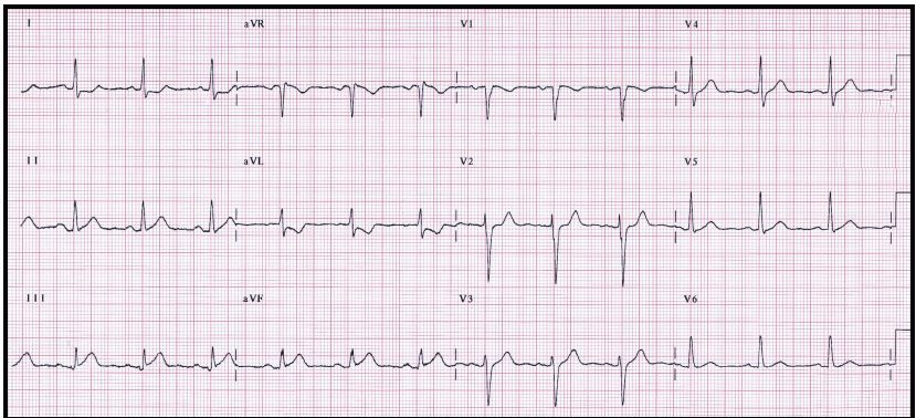
Figure 1.

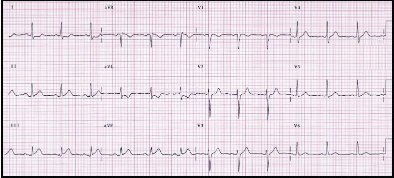
Figure 2a.

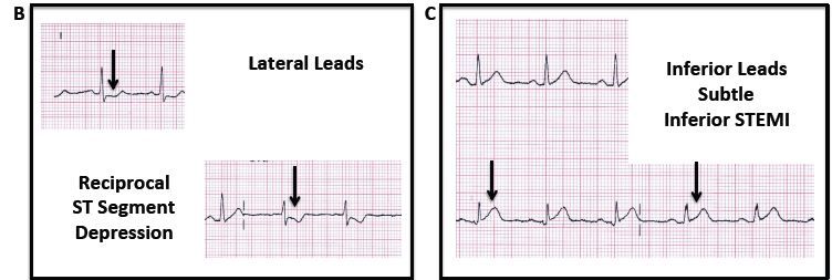
Figures 2b and 2c.

A 57-year-old woman with a history of hypertension, diabetes mellitus, and angina presented to the emergency department with chest pain. She noted the onset of chest pain approximately 2 hours before arrival at the ED; the pain was associated with dyspnea, emesis, and diaphoresis.
On examination, she was alert and oriented and in significant distress; significant diaphoresis was noted. Vital signs were: blood pressure, 105/64 mm Hg; pulse, approximately 70 beats/min; respiratory rate, 24 breaths/min; temperature of 37.4°C (99°F); and oxygen saturation of 92% on room air. The examination was otherwise normal. Results of a 12-lead ECG are seen in Figure 1.
Which ECG diagnostic tool or finding is most appropriate to confirm the diagnosis of STEMI in this patient?
A. Serial ECG analysis
B. Additional ECG leads
C. ST-segment trend monitoring
D. Reciprocal change
Answer: D. Reciprocal change
Discussion
Reciprocal ST-segment depression, also known as reciprocal change, is defined as ST-segment depression in leads separate and distinct from leads that reflect ST-segment elevation (note ST-segment depression in leads I and AVL in Figure 2B); in other words, the concept of reciprocal change cannot be used if ST-segment elevation is not present. The concept of reciprocal change cannot be used in patients with abnormal intraventricular conduction, including left bundle-branch block, ventricular paced rhythm and, to a lesser extent, left ventricular hypertrophy via voltage criteria.
The morphology of the depressed ST segment is either horizontal or downsloping; it need only be present in a single lead, but frequently is present in numerous leads. Reciprocal change is seen in approximately 75% of patients with inferior wall AMIs; in 30% of patients with anterior wall myocardial infarctions, and frequently in lateral STEMI.
Reciprocal change is an important ECG concept to consider for two reasons. First, it identifies patients with a high-risk ACS presentation. Reciprocal change in the setting of STEMI identifies a patient with an increased likelihood of cardiovascular complication (heart block, malignant ventricular dysrhythmia, cardiogenic shock) and poor outcome (significant left ventricular dysfunction, death). Second, the presence of reciprocal change is strong confirmatory evidence that STEMI is present and has both very high specificity and a positive predictive value greater than 90%.1,2 With ECG presentations that are straightforward, reciprocal changes does not assist with ECG diagnosis; in more subtle presentations, such as seen in our patient (Figures 2A and 2C), reciprocal change (Figure 2B) can aid in making the ECG diagnosis.
Case Conclusion
The ECG demonstrated subtle inferior STEMI; the presence of reciprocal change in the lateral leads strongly supports the presence of STEMI. The patient was taken immediately to the catheterization laboratory for urgent PCI. A proximal right coronary artery lesion was successfully stented. Serum troponin values demonstrated the rise and fall pattern characteristic of acute myocardial infarction. The patient was discharged home on hospital day 3 in good health.
Teaching Points
- Reciprocal change is a very important ECG finding, not only supporting the diagnosis of STEMI but also indicating a high-risk patient.
- Reciprocal change is defined as ST-segment depression occurring on an ECG which also has ST-segment elevation in at least 2 leads in a single anatomic segment.
- The concept of reciprocal change cannot be used in patients who have the following patterns on ECG: left bundle-branch block, right bundle-branch block, right ventricular paced rhythm from an implanted pacemaker, and left ventricular hypertrophy via voltage criteria with strain.
References:
- Brady WJ, Perron AD, Martin ML, et al. Electrocardiographic ST segment elevation in emergency department chest pain center patients: etiology responsible for the ST segment abnormality.Am J Emerg Med. 2001;19:25-28.
- Otto LA, Aufderheide TP. Evaluation of ST segment elevation criteria for the prehospital electrocardiographic diagnosis of acute myocardial infarction. Ann Emerg Med. 1994;23:17-24.
