- Clinical Technology
- Adult Immunization
- Hepatology
- Pediatric Immunization
- Screening
- Psychiatry
- Allergy
- Women's Health
- Cardiology
- Pediatrics
- Dermatology
- Endocrinology
- Pain Management
- Gastroenterology
- Infectious Disease
- Obesity Medicine
- Rheumatology
- Nephrology
- Neurology
- Pulmonology
Pyoderma Gangrenosum: An Uncommon Cause of Cutaneous Ulceration
The lesions may mimic infectious, vascular, inflammatory and other ulcers; they do not respond to antibiotics and debridement often exacerbates the ulcer.
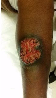
Figure 1. Lesion on admission.

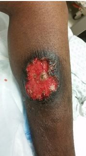
Figure 2. Lesion on Day 3.

Pyoderma gangrenosum presenting as lower limb lesions may be misdiagnosed as infected or other types of ulcers. Diagnosis is one of exclusion and physicians should be highly suspicious with concerning history and physical examination. Despite concern for pathergy, biopsy should be performed to rule out other disorders.
Case Report
A 50-year-old woman with history of hepatitis C presented to the hospital complaining of an expanding, painful, ulcerative lesion on her left leg. Patient reported a small “boil” at the center of the area approximately one month earlier. She squeezed the initial lesion and treated it with topical Neosporin. The lesion progressively expanded and she presented to a local hospital two weeks later. She was prescribed antibiotics and given instructions for daily gauze wrappings.
On presentation to our hospital, a 4.5 x 5.1-cm ulceration over the left leg was noted with undermined edge, clean base, and surrounding dark indurated skin (Figure 1; click image to enlarge). Patient was afebrile, without leukocytosis and special tests for antineutrophil cytoplasmic antibody (ANCA) and rheumatoid factor (RF) were sent. Based on history and presentation, clinical suspicion for ulcerative pyoderma gangrenosum was high. Hepatology was consulted (based on hepatitis C history) and a decision was made, based on disease severity (lesion size) and associated pain, to immediately initiate treatment with systemic prednisone, 1mg/kg daily for a dose of 60 mg. A xeroform nonadherent wound dressing was placed. Dermatology service was consulted and biopsy performed.
The patient’s pain level had improved and the appearance of the lesion was stable on day 3 (Figure 2; click image to enlarge) and she was discharged home with clinic follow up. Biopsy revealed chronic ulcer with mixed inflammation without diagnostic features of a vasculitis or neoplastic process. Special stains for microorganisms were negative.
Discussion
Pyoderma gangrenosum is a rare neutrophilic dermatosis with an estimated incidence of three to ten cases per million per year.1 It is a painful skin ulceration which may mimic infectious, vascular, inflammatory and other ulcers, eg, cryoglubulinemia.2,3 There are four basic types of pyoderma gangrenosum: ulcerative, bullous, pustular, and superficial granulomatous. Pyoderma gangrenosum is a diagnosis of exclusion, however diagnostic criteria have been proposed.4 Our patient initially met one major and one minor criterion with rapid progression of painful cutaneous ulcer and history suggestive of pathergy, respectively. Two other minor criteria were then met with histopathologic findings on biopsy and rapid treatment response to glucocorticoid therapy.
Treatment is based on clinical assessment of disease severity. For very mild disease, limited to small superficial ulcerations, a trial of potent topical corticosteroids is warranted. In cases where there is failure to respond to local therapy or initial presentation with large or disseminated ulcerations, systemic corticosteroids are recommended as they are associated with rapid improvement in pain and stabilization of lesions.3,5 Oral corticosteroids are recommended for at least several weeks.3
References:
Ruocco E, Sangiuliano S, Gravina AG, Miranda A, Nicoletti G. Pyoderma gangrenosum: an updated review. J Eur Acad Dermatol Venereol. 2009;23:1008-1017. http://onlinelibrary.wiley.com/doi/10.1111/j.1468-3083.2009.03199.x/abstract
Haines S, Hickey B, Wilson C. Pyoderma gangrenosum mimicking an infected foot. ANZ J Surg. 2015 Mar 12. doi: 10.1111/ans.13042 http://onlinelibrary.wiley.com/doi/10.1111/ans.13042/abstract
Panuncialman J, Falanga V. Unusual causes of cutaneous ulceration. Surg Clin North Am. 2010;90:1161-1180. http://www.ncbi.nlm.nih.gov/pmc/articles/PMC2991050/ Free full text
Su WP, Davis MD, Weenig RH, Powell FC, Perry HO. Pyoderma gangrenosum: clinicopathologic correlation and proposed diagnostic criteria. Int J Dermatol. 2004;43:790-800. http://onlinelibrary.wiley.com/doi/10.1111/j.1365-4632.2004.02128.x/abstract
Reichrath J, Bens G, Bonowitz A, Tilgen W. Treatment recommendations for pyoderma gangrenosum: an evidence-based review of the literature based on more than 350 patients. J Am Acad Dermatol. 2005;53:273-283. http://www.jaad.org/article/S0190-9622%2804%2902737-9/abstract
