- Clinical Technology
- Adult Immunization
- Hepatology
- Pediatric Immunization
- Screening
- Psychiatry
- Allergy
- Women's Health
- Cardiology
- Pediatrics
- Dermatology
- Endocrinology
- Pain Management
- Gastroenterology
- Infectious Disease
- Obesity Medicine
- Rheumatology
- Nephrology
- Neurology
- Pulmonology
Pseudosac with Ectopic Pregnancy
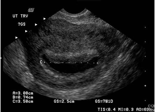
Results of a pelvic sonogram reveal the etiology of lower abdominal pain and vaginal bleeding in a 26-year-old woman.
Figure. Transvaginal sonogram.

A 26-year-old woman with a history of asthma, appendectomy, and ovarian cyst presents to the ED with lower abdominal pain accompanied by vaginal bleeding of 2 days' duration. She denies any recent fever, vomiting, vaginal discharge besides blood, or other associated symptoms. She states that she is sexually active with her husband and they use condoms for contraception. She thinks her last menstrual period was about 5 weeks ago but she is often irregular and so has not performed a home pregnancy test.
Her vital signs are all normal. Her head and neck examination is unremarkable and her lungs are clear without wheezing. The rest of her physical examination is essentially normal except for mild suprapubic tenderness. The pelvic examination was deferred as it was considered to be low yield.
A urine analysis and complete blood count (CBC) are performed with results that are normal but, perhaps not surprisingly, her pregnancy test comes back positive. A pelvic sonogram is completed by the ultrasound technologist. One of the transvaginal images is shown in the Figure.
What does the sonogram show?
What is your diagnosis? Answer: Pseudosac with likely ectopic pregnancy
Discussion
The sonogram shows a sac in the uterus but this is not a normal gestational sac or a normal pregnancy. The presence of a pseudosac rather than a true gestational sac means that the pregnancy is likely elsewhere, or ectopic. Pseudosacs occur in about 10% of ectopic pregnancies and can lead the ultrasound technician, the radiologist, or the treating provider astray if they are identified as a sign of a normal pregnancy and not seen for what they truly are-a potential red herring.
As opposed to a normal gestational sac, a pseudosac is typically located centrally and may fill the entire endometrial cavity and it also lacks the “double ring” sign. True gestational sacs will usually be slightly off-center and will have a detectable double decidual sign. True sacs also will eventually develop an internal yolk sac later followed by an embryo, but pseudosacs will have neither.
Ectopic pregnancy classically presents with steady unilateral lower abdominal pain and amenorrhea. If the ectopic ruptures and particularly if internal bleeding is significant, the abdominal pain may be generalized with abdominal distension and syncope or near syncope. There are many atypical presentations, however, to be aware of. The pain may be crampy or midline similar to that of a threatened miscarriage and there may even be vaginal bleeding, though it is usually scant. In rare instances, pain may even be absent.
Testing includes a pregnancy test, blood typing, a CBC, and a pelvic ultrasound. If the patient is unstable the ultrasound should be performed at the bedside. Classic ultrasound findings include an abnormal adnexal mass and an empty uterus. If the ectopic has ruptured there may be free fluid or clotted blood in the pelvis and/or abdomen. Though a small amount of free fluid in the pelvis may be considered “physiologic” in the non-pregnant female, it should be considered abnormal in the pregnant female. Unfortunately the ultrasound can be normal in an early ectopic pregnancy and a pseudosac, as mentioned, is a potential pitfall that can mimic a normal pregnancy.
Management for ectopic pregnancy depends on the clinical status and findings. A ruptured ectopic pregnancy requires immediate surgery. Unruptured ectopics may be treated surgically or with methotrexate and close follow-up. Patients with inconclusive findings are usually sent home with 48 hour follow-up to recheck the beta-HCG for a rise or fall but are also given precautions to return immediately for any worsening symptoms, especially upper abdominal or shoulder pain, abdominal distension, weakness, or fainting. See the Table for more details
Table.
ECTOPIC PREGNANCY
