- Clinical Technology
- Adult Immunization
- Hepatology
- Pediatric Immunization
- Screening
- Psychiatry
- Allergy
- Women's Health
- Cardiology
- Pediatrics
- Dermatology
- Endocrinology
- Pain Management
- Gastroenterology
- Infectious Disease
- Obesity Medicine
- Rheumatology
- Nephrology
- Neurology
- Pulmonology
Pruritic Scrotal Lesions

A 30-year-old man presented with multiple, mildly pruritic, nonpainful lesions on his scrotum of at least 3 months’ duration. He denied any penile discharge, dysuria, testicular swelling, fever, or other systemic symptoms. He reported a history of similar lesions on 2 occasions, which were removed surgically in another country. He was unaware of the diagnosis. He had no other significant past medical history and no current sick contacts.
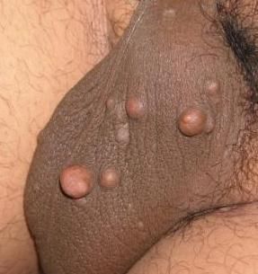
Physical examination revealed multiple firm, subcutaneous nodules bilaterally in the scrotum. No erythema or discharge was present (Figure). The lesions were non-tender and superficial. There was no inguinal lymphadenopathy. There were no other lesions noted on the body and the rest of the examination was unremarkable.
What's Your Diagnosis?
A. Epidermal inclusion cyst
B. Steatocystoma
C. Calcinosis cutis
D. Lipoma
E. Lymphangioma circumscriptum
Answer: C-Calcinosis cutis of the scrotum
Scrotal calcinosis is a rare, benign, local process characterized by multiple, painless, hard nodules that occurs in the absence of systemic manifestations. It is often referred to as idiopathic calcified nodules of the scrotum.
In this case, histologic evaluation of a biopsied lesion showed aggregates of amorphous calcified debris surrounded by a histiocytic and giant cell inflammatory response within the dermis. Numerous mitoses were present with surrounding fibrosis. These findings were consistent with a diagnosis of calcified nodules.
The benign nature of the lesions was discussed with the patient and he was scheduled for a follow up visit for excision. He did not return, however.
Discussion
The majority of patients with calcinosis cutis of the scrotum present in childhood or young adulthood, typically with solitary or multiple, calcified, painless papules or nodules. The nodules are frequently asymptomatic, but breakdown of the lesions may cause drainage of a chalky white material, itching, or a feeling of heaviness and discomfort in the scrotum.
Definitive diagnosis is made by biopsy. Histologic examination reveals extensive deposition of calcium in the dermis, which may be surrounded by histiocytes and an inflammatory giant cell reaction.1The exact etiology is unknown, but theories include idiopathic calcification within normal scrotal collagen,2 dystrophic calcification of inflamed scrotal epidermoid cysts,1 or trauma to and calcification of eccrine glands3 or dartos muscle.4 The largest case report to date, (20 patients)5 concluded that dystrophic calcification is the most likely cause.
Differential Diagnosis
Since clinical findings are nonspecific and definitive diagnosis is based on histology, the differential is broad and includes epidermal inclusion cyst, steatocystoma, lipoma, and lymphangioma circumscriptum.
Epidermoid inclusion cysts are the most common cutaneous cysts. They are the result of the implantation of epidermal elements in the dermis and. appear as flesh–colored-to-yellowish, firm, round nodules of variable size. A central pore or punctum may be present. They are rare in the genital region. They are benign lesions; however, very rare cases of associated malignancies have been reported.6
Steatocystoma is a rare, benign disorder of the pilosebaceous unit characterized by the development of numerous sebum-containing dermal cysts. It is most commonly an autosomal dominant inherited disorder, though sporadic cases can occur.7 It is characterized by multiple, smooth, flesh-to-yellow–colored cysts that are usually non-tender and asymptomatic.
Lipomas are the most common soft tissue tumors. They are benign, slow-growing, and fatty. They are soft, lobulated subcutaneous masses enclosed by a thin, fibrous capsule. Lipomas must be differentiated from other masses or tumors, such as liposarcomas. They should be removed if they cause cosmetic concern, are symptomatic or enlarging, or if they are greater than 5 cm.
Lymphangioma circumscriptum is the most common form of cutaneous lymphangioma. Lymphagiomas represent a congenital proliferation of lymphatic vessels. Lymphagioma circumscriptum arises in infancy, and is benign. Clinical findings consist of multiple, clear vesicles that may be pink, red, or black.
Management
Observation is appropriate if scrotal calcinosis is asymptomatic. Treatment is limited to surgical excision of the affected part of the scrotal wall, with scrotal reconstruction if a large area is involved. This is generally curative and reoccurrence is rare.
References:
References
1. Swinehart JM, Golitz LE. Scrotal calcinosis. Dystrophic calcification of epidermoid cysts. Arch Dermatol. 1982;118:985-988.
2. Shapiro L, Platt N, Torres-RodrÃguez VM. Idiopathic calcinosis of the scrotum. Arch Dermatol. 1970;102:199-204.
3. Dare AJ, Axelsen RA. Scrotal calcinosis: origin from dystrophic calcification of eccrine duct milia. J Cutan Pathol. 1988;15:142-149. 4. King DT, Brosman S, Hirose FM, Gillespie LM. Idiopathic calcinosis of scrotum. Urology. 1979;14:92-94.
5. Shah V, Shet T. Scrotal calcinosis results from calcification of cysts derived from hair follicles: a series of 20 cases evaluating the spectrum of changes resulting in scrotal calcinosis. Am J Dermatopathol. 2007;29:172-175.
6. Aloi F, Tomasini C, Pippione M. Mycosis fungoides and eruptive epidermoid cysts: a unique response of follicular and eccrine structures. Dermatology. 1993;187:273-277.
7. Cho S, Chang SE, Choi JH, et al. Clinical and histologic features of 64 cases of steatocystoma multiplex. J Dermatol. 2002;29:152-156.
