Primary Biliary Cirrhosis
A 50-year-old woman had a 6-month history of severe generalized itchiness and fatigability. There was no associated fever, abdominal pain, or joint pain. A cholecystectomy had been performed 20 years earlier. She had no family history of hypercholesterolemia or liver disease.
A 50-year-old woman had a 6-month history of severe generalized itchiness and fatigability. There was no associated fever, abdominal pain, or joint pain. A cholecystectomy had been performed 20 years earlier. She had no family history of hypercholesterolemia or liver disease.

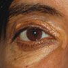
Figure 1

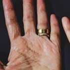
Figure 2

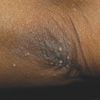
Figure 3
The patient had xanthelasma palpebrarum (Figure 1). Xanthomas were noted on the palms and elbows (Figure 2) as well as the nape and left knee. The liver was 2 cm below the costal margin, firm, non-nodular, and nontender, with a span of 10 cm. There was no splenomegaly.
Laboratory results showed markedly elevated levels of serum total cholesterol, 941.70 mg/dL; low-density lipoprotein (LDL), 890.73 mg/dL; alkaline phosphatase (ALP), 1301 U/L; alanine aminotransferase (ALT), 138 U/L; albumin, 2.2 g/L; and direct bilirubin, 2.40 mg/dL. The antimitochondrial antibody (AMA) titer was greater than 1:640. These findings suggested primary biliary cirrhosis (PBC).
Results of abdominal ultrasonography, CT, and MRI were normal. The patient was treated with ursodiol (15 mg/kg/d in 3 divided doses).
BILIARY CIRRHOSIS: AN OVERVIEW
Biliary cirrhosis is an autoimmune liver disorder that generally results from injury to or prolonged obstruction of either the intrahepatic or extrahepatic biliary system. It also may be caused by impaired biliary excretion or destruction of small intrahepatic bile ducts and canals of Hering with progressive fibrosis.1,2
PBC is characterized by chronic inflammation as well as fibrous obliteration of the intrahepatic bile ducts; the disease is progressive and can lead to liver damage and ultimately liver failure. In contrast, secondary biliary cirrhosis is defined as pro-longed obstruction of the larger extrahepatic ducts. PBC affects all races, although it is most common in whites.3 The prevalence ranges from 19 to 240 cases per million.4 The female to male ratio is 10 to 1.3 Ninety percent of affected women are between 35 and 60 years of age.2
PATHOGENESIS
Although the precise cause is unknown, both environmental and genetic factors may play a role in the pathogenesis of PBC.5 The condition is associated with automimmune disorders such as the CREST syndrome (calcinosis, Raynaud phenomenon, esophageal dysmotility, sclerodactyly, and telangiectasia), Sjögren syndrome, autoimmune thyroiditis, rheumatoid arthritis, pernicious anemia, type 1 diabetes mellitus, type 1 renal tubular acidosis, and IgA deficiency. PBC has also been reported as an extraintestinal manifestation of celiac disease.4
IgG AMA are detected in more than 90% of patients with PBC.2 These antibodies are directed against the pyruvate dehydrogenase and 2-oxo-acid enzymes in the mitochondria. In addition, a number of class II major histocompatibility loci, including DR8, have been observed in patients with PBC.
CLINICAL FEATURES
About 40% of patients are asymptomatic at the time of diagnosis. Pruritus and fatigue are the usual presenting symptoms. Pruritus can be generalized or isolated to the palms and soles2; typically the onset is insidious. The severity of fatigue is independent of the extent of the hepatic disease. Some patients may have anorexia, nausea, and right upper quadrant abdominal pain.
As the disease progresses, jaundice develops. Affected patients may have hepatomegaly, splenomegaly, hyperpigmentation, and clubbing of the fingers. Steatorrhea and malabsorption of lipid-soluble vitamins may occur because of deficiency of bile salts in the intestine and pancreatic exocrine inefficiency.6 Subcutaneous deposition of lipoprotein around the eyes (xanthelasma palpebrarum) and over joints and tendons (xanthoma) may also occur. Other clinical features include bone tenderness, glossitis, dermatitis, ascites, and ecchymosis.2
DIAGNOSIS
Blood cell counts are usually normal in the early stages of the disease.2 PBC is typically diagnosed at a presymptomatic stage based on the results of a routine liver biochemistry screening that show marked elevation of serum ALP (usually 3 to 4 times the upper limit of normal), ALT, aspartate aminotransferase (AST), and g-glutamyl transpeptidase. The diagnosis can be confirmed by the detection of AMA in the serum; this test is fairly specific and sensitive.6
Patients with PBC commonly have elevated levels of serum bile acid and serum IgM. Hypoalbuminemia, hyperbilirubinemia, and a prolonged prothrombin time-international normalized ratio are associated with poor prognosis. Tests for rheumatoid factor, anti-smooth muscle antibodies, antinuclear antibodies, and thyroid antibodies may be positive. An elevated antitransglutaminase antibody titer indicates associated celiac disease.
A cholestatic pattern of liver disease and markedly elevated cholesterol levels are hallmark features of PBC on laboratory blood work. The serum cholesterol level is high in more than half of patients and may even exceed 1003.86 mg/dL.7,8 LDL and very low-density lipoprotein levels are typically only mildly elevated, whereas high-density lipoprotein (HDL) levels are markedly increased.9
References:
REFERENCES:
1.
Washington MK. Autoimmune liver disease: overlap and outliers.
Mod Pathol.
2007;20(suppl 1):S15-S30.
2.
Chung RT, Podolsky DK. Cirrhosis and its complications. In: Kasper DL, Braunwald E, Fauci AS, et al, eds.
Harrison's Principles of Internal Medicine
. 16th ed. New York: McGraw-Hill; 2005:1857-1873.
3.
Jones DE. Pathogenesis of primary biliary cirrhosis.
Gut.
2007 Jul 19; [Epub ahead of print].
4.
Duggan JM, Duggan AE. Systematic review: the liver in coeliac disease.
Aliment Pharmacol Ther
. 2005;21:515-518.
5.
Lazaridis KN, Juran BD, Boe GM, et al. Increased prevalence of antimitochondrial antibodies in first-degree relatives of patients with primary biliary cirrhosis.
Hepatology
. 2007;46:785-792.
6.
Leuschner U. Primary biliary cirrhosis--presentation and diagnosis.
Clin Liver Dis
. 2003;7:741-758.
7.
Sherlock S, Scheuer PJ. The presentation and diagnosis of 100 patients with primary biliary cirrhosis.
N Engl J Med
. 1973;289:674-678.
8.
Iannuzzi C, Cozzolino G, Negro G. Elective cho- lecystectomy in selected cirrhotic patients.
Acta Chir Belg.
1993;93:147-150.
9.
Jahn CE, Schaefer EJ, Taam LA, et al. Lipoprotein abnormalities in primary biliary cirrhosis. Association with hepatic lipase inhibition as well as altered cholesterol esterification.
Gastroenterology
. 1985;89: 1266-1278.
10.
Blackburn P, Freeston M, Baker CR, et al. The role of psychological factors in the fatigue of primary biliary cirrhosis.
Liver Int
. 2007;27: 654-661.
11.
Ishibashi H, Komori A, Shimoda S, Gershwin ME. Guidelines for therapy of autoimmune liver disease.
Semin Liver Dis
. 2007;27:214-226.
12.
Crippin JS, Lindor KD, Jorgensen R, et al. Hypercholesterolemia and atherosclerosis in primary biliary cirrhosis: what is the risk?
Hepatology.
1992; 15:858-862.
13.
Poupon R, Chrétien Y, Poupon RE, et al. Is ursodeoxycholic acid an effective treatment for primary biliary cirrhosis?
Lancet.
1987;1:834-836.
14.
Corpechot C, Carrat F, Bahr A, et al. The effect of ursodeoxycholic acid therapy on the natural course of primary biliary cirrhosis.
Gastroenterology.
2005; 128:297-303.
15.
Gong Y, Huang Z, Christensen E, Gluud C. Ursodeoxycholic acid for patients with primary biliary cirrhosis: an updated systematic review and meta-analysis of randomized clinical trials using Bayesian approach as sensitivity analyses.
Am J Gastroenterol
. 2007;102:1799-1807.
16.
Burroughs AK, Leandro G, Goulis J. Urso- deooxycholic acid for primary biliary cirrhosis.
J Hepatol
. 2001;34:352-353.
17.
Metcalf JV, Bhopal RS, Gray J, et al. Incidence and prevalence of primary biliary cirrhosis in the city of Newcastle upon Tyne, England.
Int J Epidemiol.
1997;26:830-836.
Oral PCSK9 Inhibitor Enlicitide Meets All End Points in Phase 3 Hypercholesterolemia Trial
September 2nd 2025At week 24, patients receiving once-daily enlicitide demonstrated statistically significant and clinically meaningful reductions in low-density lipoprotein cholesterol compared with placebo.
