- Clinical Technology
- Adult Immunization
- Hepatology
- Pediatric Immunization
- Screening
- Psychiatry
- Allergy
- Women's Health
- Cardiology
- Pediatrics
- Dermatology
- Endocrinology
- Pain Management
- Gastroenterology
- Infectious Disease
- Obesity Medicine
- Rheumatology
- Nephrology
- Neurology
- Pulmonology
Photo Dx: Achilles Tendon Rupture? Tendonitis? Bursitis? Something Else?
Does an Achille’s tendon rupture, tendonitis, bursitis, or something else underlie sudden heel and lower leg pain?

HISTORY
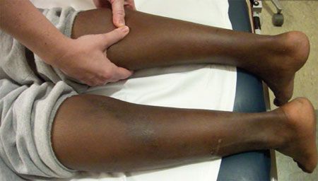
A 39-year-old man with right heel and lower leg pain comes to your office. The pain began last night during a basketball game. Another player had jumped and inadvertently landed on the back of the patient’s lower leg.
The patient is able to ambulate, although with difficulty because of the pain. He had no pain at the site before this injury, and he had never injured the right leg or foot in the past. He denies hearing a pop during the incident. No abrasions or ecchymoses are present in the area of the injury. While playing basketball, he was wearing sneakers he had owned for several months. His past medical history is significant only for depression, for which he takes citalopram.
PHYSICAL EXAMINATION
The patient is in no acute distress. There is an obvious asymmetry when the distal, posterior lower extremities are compared. The right lower extremity has diffuse edema, and a palpable groove proximal to the heel is evident. The area is tender. A Thompson test is performed bilaterally, with some flexion of the right foot but less flexion than the left foot.
WHAT’S YOUR DIAGNOSIS?
ANSWER: ACHILLES TENDON RUPTURE
This patient has an Achilles tendon rupture. Tears usually occur in an avascular area 2 to 6 cm proximal to the calcaneal insertion. Causes of rupture include high-energy disruption, degenerative changes, and mechanical imbalance. Risk factors for rupture include corticosteroid or quinolone use, chronic disease (eg, rheumatoid arthritis, systemic lupus erythematosus, and diabetes mellitus), and poor footwear.1
DIAGNOSIS
The diagnosis is typically clinical. The most used adjunct to clinical examination is the Thompson test. Also known as the Simmonds test or calf squeeze, this test was first described in 1957.2 The patient is placed either prone with feet hanging off the examination table or with knees flexed and legs hanging off the table. The examiner squeezes both calves. If the tendon is intact, squeezing produces plantar flexion. The test is considered positive if the affected foot remains neutral or has minimal plantar flexion compared with the unaffected foot.
However, false-negatives are seen with the Thompson test. An intact plantaris muscle can produce plantar flexion. If there is a delay in seeking medical attention, hematoma and inflammation will infiltrate the affected area and also potentially create plantar flexion.2 About 20% to 25% of ruptures are missed by the first examining physician.1
Ultrasonography is increasingly used in the acute setting to diagnose Achilles tendon rupture when a patient presents with confounding physical findings.3 This study also allows a complete tear to be distinguished from a partial tear, which cannot be discerned on physical examination. Although MRI is another modality that is frequently used in this setting, ultrasonography provides rapid dynamic imaging at the bedside and is less expensive than MRI. However, the need for an experienced operator may preclude the routine use of ultrasonography in some areas.
TREATMENT
Treatment involves referral to an orthopedic surgeon and conservative or operative management. Conservative therapy includes the use of a walking boot for 4 weeks with weight bearing as tolerated, then weaning from the boot. Although surgery is more invasive, the downfall of supportive care is a higher rerupture rate compared with surgical intervention.1
This patient underwent definitive surgical management. At his postoperative visit several weeks later, he was doing well.
DIFFERENTIAL DIAGNOSISRetrocalcaneal bursitis is an inflammation of the retrocalcaneal bursa, located between the posterior aspect of the calcaneus and the Achilles tendon. Conservative measures are the first line of treatment and are typically successful in reducing pain and inflammation.4
Haglund deformity, also known as pump bump, is a bony enlargement at the posterior aspect of the calcaneus. The retrocalcaneal bursa overlies this normal variant of the calcaneus and becomes inflamed when tight shoes rub against the bony protuberance. This deformity is often attributed to the rigid back of high-heeled shoes.4 Supportive measures are the mainstay of treatment.
Achilles tendonitis is commonly an overuse injury and may cause small tears, leaving the tendon susceptible to rupture. It may be mistaken for retrocalcaneal bursitis; however, it can be differentiated by tenderness with palpation of the distal Achilles tendon in contrast to tenderness over the posterior aspect of the calcaneus. The symptoms of Achilles tendonitis may be acute or chronic. Treatment includes supportive measures.
Editor's note: This video shows the Thompson test being performed.
References:
REFERENCES:
1.
Maffulli N, Wong J. Rupture of the Achilles and patellar tendons.
Clin Sports Med.
2003;22:761-776.
2.
Douglas J, Kelly M, Blachut P. Clarification of the Simmonds-Thompson test for rupture of an Achilles tendon.
Can J Surg.
2009;52:E40-E41.
3.
Fessell DP, Jacobson JA. Ultrasound of the hindfoot and midfoot.
Radiol Clin North Am.
2008;46:1027-1043, vi.
4.
Stephens MM. Haglund’s deformity and retrocalcaneal bursitis.
Orthop Clin North Am.
1994;25:41-46.
