- Clinical Technology
- Adult Immunization
- Hepatology
- Pediatric Immunization
- Screening
- Psychiatry
- Allergy
- Women's Health
- Cardiology
- Pediatrics
- Dermatology
- Endocrinology
- Pain Management
- Gastroenterology
- Infectious Disease
- Obesity Medicine
- Rheumatology
- Nephrology
- Neurology
- Pulmonology
Patent Foramen Ovale and Paradoxical Stroke in a Young Cyclist
PFO can be detected in 10% to 15% of the population by transthoracic echocardiogram. Autopsy studies show a prevalence of PFO of approximately 26%.
A 42-year-old man presented to the emergency department after experiencing multiple episodes of difficulty in speaking. Over the past 24 hours, had been unable to formulate his speech for 1 to 2 minutes per episode but was aware of what he wanted to say. He had experienced a similar episode 2 weeks earlier when he lost the ability to verbalize his thoughts for nearly 5 minutes.
Three years earlier, the patient had a deep venous thrombosis (DVT) that was treated with anticoagulation. He had also sustained head trauma without loss of consciousness approximately 4 years earlier.
The patient was a competitive bicyclist and he maintained a rigorous training regimen year round. His social history was otherwise noncontributory. He denied any medication allergies and was not taking any medications on a routine basis. Review of systems was unremarkable.
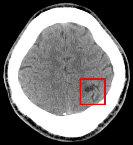
Figure 1:
CT Head showing area of old infarct in the left parietal cortex.
On examination, the patient was alert and oriented to person, place, and time. Vital signs were within normal limits. Cardiovascular and pulmonary examinations were unremarkable. The neurologic examination was grossly non-focal except for speech, which was slow and lacking in fluency, and which required significant effort. Articulation was impaired, although semantics were appropriate. The patient occasionally used filler words to complete thoughts.
Examination of the lower extremities found varicose veins, but no tenderness or edema, and a negative Homan sign bilaterally.
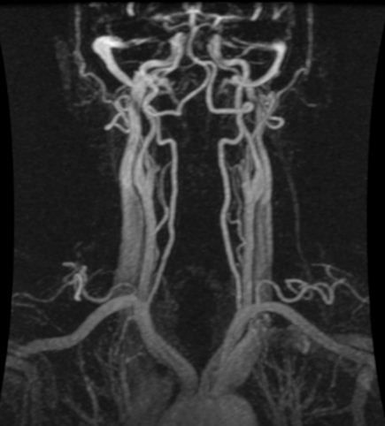
Figure 2:
MRA of neck with IV contrast.
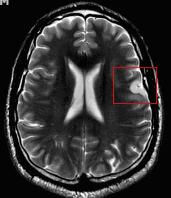
Figure 3:
MRI with IV contrast demonstrates focal diffusion restriction at the left posterior frontal lobe, which is suggestive of a subacute infarct.
A CT scan of the head showed no acute infarct but revealed evidence of an old infarct in the left parietal cortex (Figure 1). MRI of the brain without contrast suggested an acute infarct in the left frontal supraventricular white matter; these images did rule out a solid lesion or signal abnormality that might be related to seizure activity. A neurology consult was requested.
CT angiography of the head was unremarkable and MRA of the neck (Figure 2) with contrast found no signs of stenosis, thrombosis, or dissection. Subsequent MRI of the brain with contrast indicated a subacute infarct (Figure 3). An infectious disease workup was normal and ruled out focal encephalitis. MRI spectroscopy (Figure 4) verified an ischemic infarct.
Additional studies were performed to identify the cause of infarct. Doppler ultrasonography of the carotid artery showed no blockage and results of a transthoracic echocardiogram were within normal limits. Cardiolipin IgG and IgM antibody levels were within normal limits, which ruled out antiphospholipid syndrome. Results of a lipid panel also were within normal limits, which further reduced the possibility of atherosclerosis in the carotid arteries.4 A workup for vasculitis also was negative.
The patient then underwent transesophageal echocardiography, which showed a patent foramen ovale (PFO) (Figure 5). Lower extremity venous Doppler ultrasonography was negative for DVT. Anticoagulation was initiated with enoxaparin sodium, and the patient was discharged with a plan for outpatient closure of PFO. By the time of discharge, the patient’s speech had returned to baseline.
Because of our patient’s history of DVT, anticoagulation with enoxaparin and warfarin was prescribed. After 6 months of anticoagulation therapy, he underwent an elective procedure to close the PFO. Anticoagulation therapy was discontinued after the procedure. He has been in follow-up care and has had no recurrence of original symptoms and has experienced no thrombotic events.
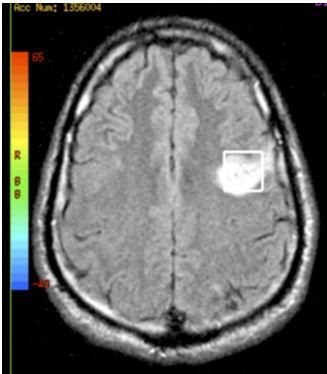
Figure 4:
A single Voxel MRI spectroscopy taken during admission revealing an ischemic infarct.
Discussion
The majority (90%) of strokes that occur annually in the United States are ischemic. PFO is not considered a primary cause of stroke. Cardioembolic events account for 19% and carotid disease accounts for 15% of ischemic strokes; the remainder is cryptogenic. Approximately 40% of cryptogenic ischemic strokes are attributed to PFO. PFO can be detected in 10% to 15% of the population by transthoracic echocardiogram. Autopsy studies show a prevalence of PFO of approximately 26%.1
A number of pathologic manifestations of PFO have been recently identified, including paradoxical systemic embolism. Neurologic decompression illness (eg, in divers, high-altitude pilots, and astronauts) and migraine headache with aura also have been associated with PFO but are not common findings.2-4 Most patients with PFO, however, remain asymptomatic because under normal physiologic conditions, PFO allows a small amount of left-to-right shunting without causing significant hemodynamic change.3 consequently, neurologic deficits associated with PFO-such as those seen in this case-are rare.
Differential diagnoses for this patient’s presentation that were ruled out include intracranial or extracranial thrombosis; carotid dissection; and coagulopathies, such as antiphospholipid antibodies.5
Management of PFO
There are currently 3 primary options for management of PFO6:
• Medical treatment with anticoagulation or antiplatelet therapy
• Surgical closure
• Percutaneous closure
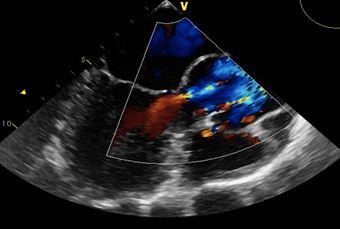
Figure 5:
A TEE shows via doppler the blood flow from the right atrium to the left atrium via a septal defect.
In a study that compared aspirin with warfarin in the prevention of recurrent cryptogenic ischemic stroke, there was no significant benefit shown in either group.7
Although there are currently no randomized clinical trials comparing these 3 treatment methods, closure of PFO appears to have clear benefits over medical therapy in select patients.5 Many studies report that transcatheter percutaneous PFO closure is safe and effective, with efficacy that ranges from 86% to 100%.7 The procedure is simple, requires only overnight observation, and has replaced surgical closure in all but rare instances.8
References:
References:
1. Di Tullio MR. Patent foramen ovale: echocardiographic detection and clinical relevance in stroke. J Am Soc Echocardiogr. 2010;23:144-155.
2. Fazio G, Ferro G, Barbaro G, et al. Patent foramen ovale and thromboembolic complications. Curr Pharm Des. 2010;16:3497-3502.
3. Guo S, Roberts I, Missri J. Paradoxical embolism, deep vein thrombosis, pulmonary embolism in a patient with patent foramen ovale: a case report. J Med Case Rep. 2007;1:104.
4. Germonpré P, Dendale P, Unger P, Balestra C. Patent foramen ovale and decompression sickness in sports divers. J Appl Physiol. 1998;84:1622-1626.
5. APASS Investigators. Antiphospholipid antibodies and subsequent thrombo-occlusive events in patients with ischemic stroke. JAMA. 2004;291:576-584.
6. Bailey CE, Allaqaband S, Bajwa TK. Current management of patients with patent foramen ovale and cryptogenic stroke: our experience and review of the literature. WMJ. 2004;103:32-36.
7. Mohr JP, Thompson JL, Lazar RM, et al. A comparison of warfarin and aspirin for the prevention of recurrent ischemic stroke. N Engl J Med. 2001;345:1444-1451.
8. Martin F, Sanchez PL, Doherty E, et al. Percutaneous transcatheter closure of patent foramen ovale in patients with paradoxical embolism. Circulation. 2002;106:1121-1126.
