- Clinical Technology
- Adult Immunization
- Hepatology
- Pediatric Immunization
- Screening
- Psychiatry
- Allergy
- Women's Health
- Cardiology
- Pediatrics
- Dermatology
- Endocrinology
- Pain Management
- Gastroenterology
- Infectious Disease
- Obesity Medicine
- Rheumatology
- Nephrology
- Neurology
- Pulmonology
Parotid Gland Deformities in HIV Seropositive Patients: The Best Choice for Cosmetic Control
More than half of people with HIV infection in the United States develop head and neck lesions. Common among these is enlargement of the parotid gland, which causes disfigurement and therefore distress. This review discusses the evidence for radiation treatment as the best option, as well as the dangers of choosing the wrong treatment for this benign comorbidity of HIV-positive status.
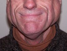
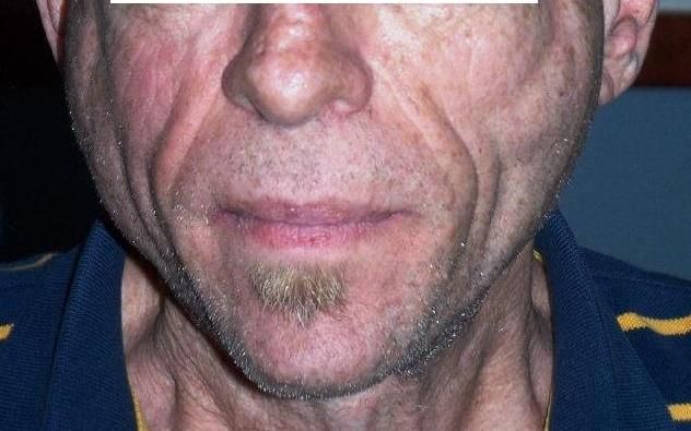
Among the 1.2 million people in the United States who are living with HIV infection (and the approximately 50,000 more who become infected with HIV each year,1 head and neck lesions associated with HIV occur in more than 50%.2 Common among these problems is enlargement of the parotid gland, usually secondary to the development of benign lymphoepithelial cysts (BLEC) within the parotid gland.

Also known as benign parotid hypertrophy or lymphoepithlioma, BLEC of the parotid gland burdens the affected patient with physical disfigurement and cosmetic distress.3 BLEC of the parotid gland has been estimated to occur in up to 6% and 10% of HIV-positive adults and children, respectively.4,15,16 Although the first case of BLEC of the parotid gland was reported by Hildebrandt in 1895, and later confirmed by Albrecht and Artz in 1910,13, 14 this disfiguring consequence of HIV infection among long-term survivors is insufficiently recognized among medical providers who do not specialize in treatment of HIV-positive patients.

Although radiotherapy (RT) has been generally recognized as the most successful and least invasive response to this condition, patients continue to receive other treatments that are either ineffective or dangerous, at the risk of infection, salivary gland damage, and facial paralysis. Here we offer our experience from the largest case series in the United States with this condition, as well as a review of the literature.
Clinical Presentation
The most common presentation in HIV patients is salivary gland swelling.4 BLEC is characterized by bilateral swelling of the parotid gland as well as diffuse CD8 infiltration, with or without cervical lymphadenopathy.5-7. BLEC has been reported to occur in the salivary glands or their lymph nodes, the oral cavity (floor of mouth), tonsils, and thyroid gland.4, 8-12
The exact pathogenesis of BLEC is unclear, but the most common hypotheses are viral replication, proliferation of the glandular epithelium that is trapped within the lymph nodes in the parotid gland, and lymphoid proliferation causing ductal obstruction and dilation that results in cyst formation and subsequent parotid enlargement.8, 18-20 Histologically, lymphoepithelial cysts appear to arise in the parotid nodes, usually with lymphoid hyperplasia surrounding the cystic spaces. The cysts are densely infiltrated lymphoidal cells and are partially encapsulated by a fibrous capsule.18 This lymphocytic infiltration may also result in xerostomia caused by destruction of acinar tissue.21


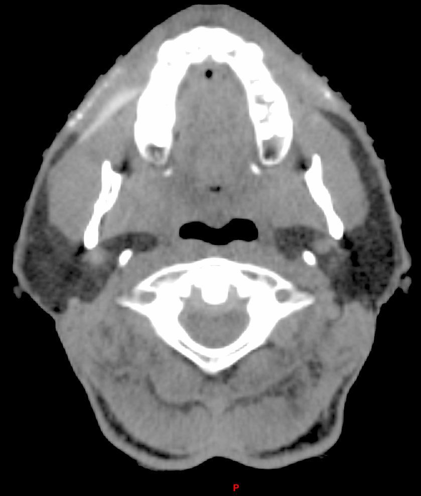
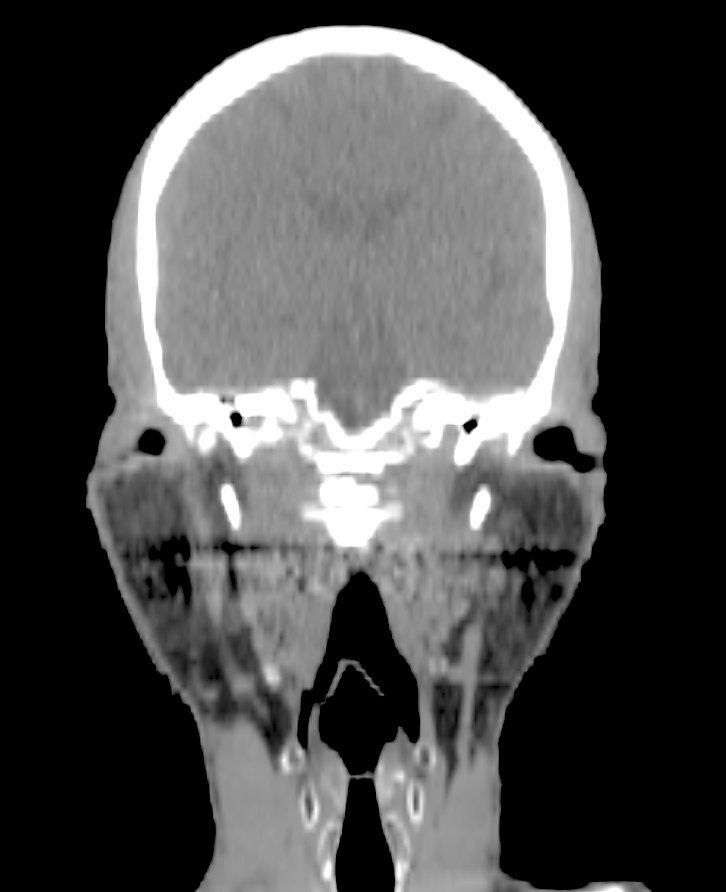
BLEC is due to multicystic, benign lymphoepithelial lesions of one or, more commonly both, parotid glands.25The classical clinical presentation is bilateral (up to 80%), with parotid swellings that are multiple (up to 90%), painless, soft, and slowly progressive in size. Unilateral parotid swellings have also been reported.4, 22 Rarely does BLEC involve any other salivary gland; however, submandibular gland involvement has been reported.8
The diagnostic evaluation consists mainly of ultrasound scan, CT, and/or MRI showing multiple thin walled cysts.4 (See CT scans above.)
Treatment Options
The treatment options for BLEC include close observation, repeat aspiration, highly active antiretroviral therapy (HAART), sclerosing therapy, RT and surgery.4 Observation of BLEC is an option due to the slowly progressing nature of the disease, but any sudden increases in gland size warrants immediate investigation and intervention.
As the parotid gland enlarges and becomes disfiguring, some patients may seek aspiration, which is a quick office-based procedure.17 The main disadvantage is that most aspirated lesions will recur within weeks to months and subsequently continue to grow.4,18 However, FNA has been shown to be an effective diagnostic and therapeutic tool for BLEC and for monitoring for the development of EBV-associated malignant B-cell lymphoma, of which these patients are at an increased risk.21
Sclerotherapy with doxycycline has also been used for the treatment of BLEC.10 Studies with short-term results have reported an average reduction in cyst size by 42% to 100%.10, 17 Although sclerotherapy is known for causing mild edema and tenderness, no serious complications (i.e., facial nerve injury, infection) have been reported.
Surgery is rarely recommended for BLEC, given the bilateral and progressive nature of the disease, and the risk of facial nerve injury. Multiple surgical procedures would be required during the course of the disease, even after a superficial parotidectomy.4
External beam RT has shown significant promise in reducing BLEC and parotid gland size.29 Some investigators have advocated RT as the standard of care for the treatment of BLEC.26-29 Goldstein et al reported that the initial response rates of BLEC to low dose RT were encouraging, but longer follow-up revealed a median duration of control of 9.5 months.26, 27 A few years later, they updated their study to report that after initial RT failure, retreatment of eight patients with low RT doses produced less response than the initial RT and did not achieve long-term local control.27 This led to cautious exploration of the need for a higher radiation dose.28, 29
In a series by Beitler et al, 24 HIV patients with BLEC were treated with a moderate RT dose and had an excellent local control after a median follow up of 2 years.28 They compared these updated results with the earlier series, finding a significant improvement in cosmetic control (p <0.02).29 They concluded that a moderate RT dose is a safe, well-tolerated procedure that appeared effective in 68% of patients.
HAART has also been found to reduce and eliminate BLEC.24 Based on our most recent and largest single institution study30 which was presented at the multidisciplinary head and neck cancer symposium (January 26-28, 2012, Phoenix, AZ), we agree with Beitler and colleagues that HAART should precede RT, that RT should subsequently be offered to those whose parotid disease is refractory or resistant to HAART. At our institution, the standard practice is: seropositive HIV confirmation, CD4 counts, CBC, viral load, tissue biopsy via FNA (mainly to confirm the diagnosis and exclude other possible causes such as lymphoma), and baseline CT scan to determine the pretreatment parotid and BLEC sizes.
For all our patients, we deliver CT based planning and three dimensional conformal radiotherapy (3D-CRT). As stated above, we recommend that HAART should precede RT. Only for those HIV-positive patients whose parotid disease is refractory or resistant to HAART do we offer RT, in order to cosmetically control the BLEC of the parotid glands.
Cautions
The potential for radiation-induced malignancy does exist, but to the best of our knowledge as a result of our extensive review of the English medical literature, like Beitler et al29 we were unable to identify a specific case that has occurred as a result of the treatment of HIV-BLEC with RT. However, all patients should be informed of this risk, as survival duration has improved with the advances in HAART.
Non-compliance with HAART and RT is an important problem in this patient population. In our recent study, we found that treatment interruptions were mainly due to psychosocial and logistic issues, rather than medical reasons. This is consistent with the literature showing that patients with anxiety and depression, both of which are fairly common in the HIV population, are more likely to experience treatment breaks. Given our data, in conjunction with those from others demonstrating that psychosocial stresses can negatively influence treatment outcomes in the HIV positive population, it seems reasonable to offer referral to counseling services for patients with this condition who are unable to adhere to the recommended course of treatment.
To date, RT is still the recommended treatment modality. Evidence to support any other option is scarce and lacks long-term follow-up data. With the low incidence of side effects, we agree with Beitler et al that RT should be considered the standard of care for those patients, for whom adverse cosmetic outcomes would detract considerably from their quality of life.29-30
References:
1. Estimated Number of New HIV Cases, CDC. US Centers for Disease Control. www.cdc.gov/hiv. November, 2011.
2. Yengopal V, Naidoo S. Do oral lesions associated with HIV affect quality of life? Oral Surg Oral Med Oral Pathol Oral Radiol Endod 2008;106(1):66â73.
3. Adedigba M, Ogunbodede E, Jeboda S, et al. Patterns of oral manifestations of HIV/AIDS among 225 Nigerian patients. OralDis 2008;14(4):341â6.
4. Dave S, Pernas F, Roy S. The benign lymphoepithelial cyst and a classification system for lymphocytic parotid gland enlargement in the pediatric HIV population. Laryngoscope 2007;117(1):106â13.
5. Mandel L, Alfi D. Drug-induced paraparotid fat deposition in patients with HIV. JAmDentAssoc 2008;139(2):152â7.
6. Mandel L, Kim D, Uy C. Parotid gland swelling in HIV diffuse infiltrative CD8 lymphocytosis syndrome. Oral Surg Oral Med Oral Pathol Oral Radiol Endod 1998;85:565â8.
7. Schrot R, Adelman H, Linden C, et al. Cystic parotid gland enlargement in HIV disease: the diffuse infiltrative lymphocytosis syndrome. JAMA 1997;278:166â7.
8. Elliott J, Oertel Y. Lymphoepithelial cysts of the salivary glands: histologic and cytologic features. AmJClinPathol 1990;93:39â42.
9. Weidner H, Geisinger K, Sterling R, et al. Benign lymphoepithelial cysts of the parotid gland: a histologic, cytologic, and ultrastructural study. AmJClinPathol 1986;85:395â401.
10. Lustig R, Lee K, Murr A, et al. Doxycycline sclerosis of benign lymphoepithelial cysts in patients infected with HIV. Laryngoscope 1988;108:1199â205.
11. Favia G, Capodiferro S, Scivetti M, et al. Multiple parotid lymphoepithelial cysts in patients with HIV infection: report of two cases. Oral Dis 2004;10:151â4.
12. Brudnicki A, Levin T, Slim M, et al. HIV-associated (non-thymic) intrathoracic lymphoepithelial cyst in a child. Pediatr Radiol 2001;31:603â5.
13. Hildebrandt O. Arch. Klin. Chir. 49: 167;1895.
14. Albrecht H, and Arts L. Frankfurt Z.f.Path.4:47;1910.
15. Morris M, Moore D, Shearer G. Bilateral multiple benign lymphoepithelial cysts of the parotid gland. Otolaryngol Head Neck Surg 1987;97:87â90.
16. Huang R, Pearlman S, Friedman W, et al. Benign cystic vs. solid lesions of the parotid gland in HIV patients. Head Neck 1991;13:522â7.
17. Echavez M, Lee K, Sooy C. Tetracycline sclerosis for treatment of benign lymphoepithelial cyst of the parotid gland in patients infected with human immunodeficiency virus. Laryngoscope 1994;104: 1499â502.
18. Mandel L, Reich R. HIV parotid gland lymphoepithelial cysts. Oral Surg Oral Med Oral Pathol 1992;74: 273â8.
19. Mandel L, Hong J. HIV-associated parotid lymphoepithelial cysts. JAmDent Assoc 1999;130(4):528â32.
20. Ihrler S, Zeitz C, Riederer A, et al. HIV-related parotid lymphoepithelial cysts. Immunohistochemistry and 3-D reconstruction of surgical and autopsy material with special reference to formal pathogenesis. Virchows Arch 1996;429:139â47.
21. Patton L, van-der-Horst C. Oral infections and other manifestations of HIV disease. Infect Dis Clin North Am 1999;13(4):879â900.
22. Al-Maawali A, Chacko A, Javad H, et al. HIV disease presenting as a unilateral parotid gland swelling. IndianJPediatr 2008;75(10):1087â8.
23. Mayer M, Haddad J. Human immunodeficiency virus infection presenting with lymphoepithelial cysts in a six-year old child. Ann Otol Rhinol Laryngol. 1996; 105:242â4.
24. Shaha A, DiMaio T, Webber C, et al. Benign lymphoepithelial lesions of the parotid. AmJSurg.1993;166:403â6.
25. Sperling N and Lin P. Parotid disease associated with human immuno deficiency virus infection. Ear Nose Throat J 1990;69:475â 477.
26. Goldstein J, Rubin J, Silver C, et al. Radiation therapy as a treatment for benign lymphoepithelial parotid cysts in patients infected with human immunodeficiencyvirus-1. Int J Radiat Oncol Biol Phys1992;23:1045â1050.
27. Beitler J, Vikram B, Silver C, et al. Low dose radiotherapy for multicystic benign lymphoepithelial lesions of the parotid gland in HIV-positive patients: Long-term results. Head & Neck 1995;17:31â35.
28. Beitler J, Smith R, Silver C, et al. Cosmetic control of parotid gland hypertrophy using radiation therapy. Aids Patient Care 1995;9:271â275.
29. Beitler J, Smith R, Brook A, et al. Benign parotid hypertrophy in HIV patients: limited late failures after external radiation. IntJRadiatOncolBiolPhys. 1999 Vol. 45, No. 2, pp. 451â455.
