- Clinical Technology
- Adult Immunization
- Hepatology
- Pediatric Immunization
- Screening
- Psychiatry
- Allergy
- Women's Health
- Cardiology
- Pediatrics
- Dermatology
- Endocrinology
- Pain Management
- Gastroenterology
- Infectious Disease
- Obesity Medicine
- Rheumatology
- Nephrology
- Neurology
- Pulmonology
Papilledema in Neurosarcoidosis
Six days ago, a 36-year-old man had noticed a dark spot in the field of vision of his left eye. Now the spot more closely resembled a line. He denied other changes in his vision and had not seen any floaters or flashing lights.

Six days ago, a 36-year-old man had noticed a dark spot in the field of vision of his left eye. Now the spot more closely resembled a line. He denied other changes in his vision and had not seen any floaters or flashing lights.
Two months earlier, the patient had swelling of the ankles and wrists. A complete workup by a rheumatologist was unremarkable except for an elevated C-reactive protein level of 2.57 mg/dL (normal, less than 0.9 mg/dL) and the finding of a mild prominence of the left hilum on his chest radiograph. Acute reactive arthritis was diagnosed, with sarcoidosis as the possible underlying cause. The patient was treated with oral prednisone, which he was still taking at the time of presentation.
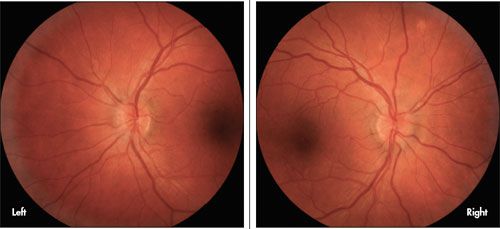
Uncorrected visual acuity was 20/20 in each eye. The pupils, motility, tonometry, and slitlamp evaluation were all normal. No deficits were found on a computerized visual field analysis. Examination of the optic discs through dilated pupils revealed bilateral papilledema, more prominent at the nasal disc margins (see photos).
Results of MRI, angiography, and venography were normal. A lumbar puncture revealed an elevated opening pressure of 250 mm H2O and lymphocytic pleocytosis. A CT scan of the chest showed mediastinal and hilar lymphadenopathy. A transbronchial biopsy demonstrated non-necrotizing granulomas, which confirmed the diagnosis of neurosarcoidosis.
Treatment with prednisone was continued, and oral methotrexate was started. At follow-up 7 weeks later, the patient's visual symptoms had resolved and the disc edema had greatly diminished.
Optic nerve dysfunction is the most common neuro-ophthalmological manifestation of sarcoidosis, and the optic nerve is said to be the second most commonly affected cranial nerve after the facial nerve.1
The optic nerve may be affected at any time during the course of the disease and may be the site of its initial presentation.2 Papilledema, as seen in this patient, may result from the elevated intracranial pressure, which is often associated with neurosarcoidosis.
References:
1. Hamilton SR. Sarcoidosis. In: Miller NR, Newman NJ, eds. Walsh & Hoyt's Clinical Neuro-Ophthalmology. 6th ed. Philadelphia: Lippincott Williams & Wilkins; 2004:3369-3427.
2. Jordan DR, Anderson RL, Nerad JA, et al. Optic nerve involvement as the initial manifestation of sarcoidosis. Can J Ophthalmol. 1988;23:232-237.
