- Clinical Technology
- Adult Immunization
- Hepatology
- Pediatric Immunization
- Screening
- Psychiatry
- Allergy
- Women's Health
- Cardiology
- Pediatrics
- Dermatology
- Endocrinology
- Pain Management
- Gastroenterology
- Infectious Disease
- Obesity Medicine
- Rheumatology
- Nephrology
- Neurology
- Pulmonology
Orbital Pseudotumor Disguised as Orbital Cellulitis and Sinusitis
A 58-year-old man with a past medical history of chronic sinus disease and hypothyroidism presented with left periorbital pain and erythema that worsened despite outpatient treatment with topical antibiotics. An outpatient CT scan showed pansinusitis and orbital stranding. The diagnosis was orbital cellulitis and sinusitis.
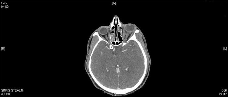
A 58-year-old man with a past medical history of chronic sinus disease and hypothyroidism presented with left periorbital pain and erythema that worsened despite outpatient treatment with topical antibiotics. An outpatient CT scan showed pansinusitis and orbital stranding (Figure 1). The diagnosis was orbital cellulitis and sinusitis.

Figure 1
.
The patient complained of blurry vision with lateral gaze, headache, pain with eye movements, foreign body sensation, and nasal congestion. He denied fevers, sick contacts, rash, or recent travel. Vital signs were stable and he was afebrile. The left eye examination was significant for ptosis, proptosis, periorbital edema, chemosis, erythema, and resistance to retropulsion (Figure 2). Visual acuity was 20/20. Pupils were equal, round, and reactive, without afferent pupillary defect. Extraocular movements showed restriction of abduction of the left eye. Intraocular pressure was within normal limits and symmetric. Findings from funduscopic examination were unremarkable.
Results of bloodwork, including blood cultures and thyroid studies, were unremarkable except for a peripheral eosinophilia of 11%. At this time, a CT showed complete pansinusitis and left orbital fat stranding.


Figure 2.
The patient was treated with IV cefepime and metronidazole. A nasal culture grew methicillin-sensitive Staphylococcus aureus. Despite antibiotics, the condition did not improve and an MRI on hospital day 6 revealed pansinusitis and orbital cellulitis. An inflammatory process could not be excluded.
On hospital day 7, the patient underwent surgical drainage. Pathology showed sinonasal mucosa with edema, inflammation, numerous eosinophils, lymphocytes, few neutrophils, and occasional plasma cells. Cultures were negative for acid-fast bacilli and fungi. He was given a short course of corticosteroids and was discharged on a 3-week regimen of oral moxifloxacin.
Figure 3.

The orbital signs had improved 1 week after discharge (Figure 3). Two weeks after discharge, however, the patient returned with increased swelling. He was advised to continue taking moxifloxacin and was given a methylprednisolone dose pack. At follow-up 1 week later, he was feeling well and his eye exam was normal.

Figure 4.
Seven weeks after discharge, and 1 month after antibiotics and corticosteroids had been discontinued, the patient returned with increased swelling and orbital signs (Figure 4). MRI revealed marked improvement of sinus disease with persistent orbital stranding and mild swelling of the lacrimal gland. Therapy with prednisone, 60 mg/d, was started. Within 2 weeks, symptoms and examination findings were normal (Figure 5). Findings from a detailed rheumatologic workup were unremarkable. Methotrexate therapy was initiated to avoid prolonged corticosteroid therapy.
Orbital inflammatory syndrome (OIS)
Also known as orbital pseudotumor, this benign inflammatory syndrome has variable clinical presentations and is often a diagnosis of exclusion.1,2 It accounts for 5% to 10% of orbital processes.3 Ocular manifestations include pain, proptosis, ptosis, periorbital edema, conjunctival injection, and erythema.3 Visual loss and ocular dysmotility can occur.2 OIS can be unilateral or bilateral.4 Symptoms can range from acute (days) to subacaute (weeks) or chronic (weeks to months).2 The presentations vary depending on the location and degree of inflammation.4 There is no racial or sex predominance, and adults are primarily affected.2

Figure 5.
In “classic orbital pseudotumor,” the cellular infiltrate seen on histology consists of inflammatory cells-mainly mature lymphocytes, admixed with plasma cells, neutrophils, eosinophils, and occasionally macrophages and histiocytes, with stromal edema-as was seen in our patient.1 Other histologic findings have been described, including sclerosing, granulomatous, vasculitic, and eosinophilic subtypes.5 Treatment is with systemic corticosteroids, tapered slowly over several months to prevent rebound.4
Orbital cellulitis and thyroid orbitopathy are the two most common mimics of orbital pseudotumor. Orbital cellulitis typically presents as acute onset of unilateral severe pain, proptosis, and restricted eye movements. It can arise from direct extension of sinusitis or orbital infection; via hematogenous spread; or from complications of eye surgery, sinus surgery, or dental work.4 One would expect systemic symptoms such as fever and leukocytosis (which were not present in our patient). Treatment is with IV antibiotics to cover common causative organisms; surgical intervention is indicated in cases of orbital abscess formation.
In our patient, imaging was consistent with pansinusitis and orbital cellulitis, prompting treatment with broad-spectrum antibiotics. Antibiotics alone elicited mild improvement; corticosteroids yielded more improvement. After antibiotics and corticosteroids were stopped, the symptoms returned; they responded to corticosteroid therapy alone, which made the diagnosis of orbital pseudotumor likely.
The presence of pansinusitis made our diagnosis more difficult. Although more commonly seen with orbital cellulitis, it has been reported in orbital pseudotumor. Earlier literature reported a possible association between paranasal sinusitis and orbital pseudotumor.6-9 Others have reported that the two are distinct clinical entities and not a direct extension of inflammation from the orbit to the sinuses.10 Orbital pseudotumor, although typically confined to the orbit, can have extraorbital extension. A series of 4 cases demonstrated 1 with maxillary sinus extension and 2 with intracranial extension.11 A recent review of 91 patients with orbital inflammatory syndrome found 6 with significant sinus inflammation (6.6%).1 Four of the 6 patients underwent sinus surgery and 1 an orbital biopsy. All had histopathology consistent with inflammation without evidence of infection or vasculitis. As in our patient, imaging demonstrated extraocular muscle enlargement and orbital fat stranding or haziness. The ipsilateral maxillary sinus was involved in all of these cases, as in our patient.
Take-home points
Consider the diagnosis of OIS in patients with orbital signs in the setting of sinusitis, especially in those whose symptoms do not improve with antibiotics alone. An association of sinusitis with orbital inflammatory syndrome cannot be proved on the basis of this case alone. However, the case demonstrates that a possible association may exist. It also depicts the difficulty in making the diagnosis of OIS when there is concomitant sinus disease.
In our patient, despite significant sinus improvement on imaging after antibiotics and surgery, the orbital symptoms and findings on imaging recurred and then improved with corticosteroids alone. This supports the diagnosis of OIS.
References1. Leibovitch I, Goldberg RA, Selva D. Paranasal sinus inflammation and non-specific orbital inflammatory syndrome: an uncommon association. Graefes Arch Clin Exp Ophthalmol. 2006;244:1391-1397.
2. Anderson J, Thomas T. Orbital pseudotumor presenting as orbital cellulitis. Can J Emerg Med. 2006;8:123-125.
3. Chaudhry IA, Shamsi FA, Arat YO, et al. Orbital pseudotumor: distinct diagnositic features and management. Middle East Afr J Ophthalmol. 2008;15:17-27.
4. Zerilli TC, Burke CL. Orbital pseudotumor after an upper respiratory infection: a comprehensive review. Optometry. 2010;81:638-646.
5. Swamy BN, McCluskey P, Nemet A, et al. Idiopathic orbital inflammatory syndrome: clinical features and treatment outcomes. Br J Ophthalmol. 2007;91:1667-1670.
6. Blodi FC, Gass JD. Inflammatory pseudotumor of the orbit. Trans Am Acad Ophthalmol Otolaryngol. 1967;71:303-323.
7. Blodi FC, Gass JD. Inflammatory pseudotumor of the orbit. Br J Ophthalmol. 1968;52:79-93.
8. Fortson JK, Shapshay SM, Weiter JJ, et al. Otolaryngologic manifestations of orbital pseudotumors. Otolaryngol Head Neck Surg. 1980;88:342-348.
9. Heersink B, Rodrigues MR, Flanagan JC. Inflammatory pseudotumor of the orbit. Ann Ophthalmol. 1977;9:17-22, 25-29.
10. Eshaghian J, Anderson RL. Sinus involvement in inflammatory obital pseudotumor. Arch Ophthalmol. 1981;99:627- 630.
11. Mahr MA, Salomao DR, Garrity JA. Inflammatory orbital pseudotumor with extension beyond the orbit. Am J Ophthalmol. 2004;138:396-400.
