- Clinical Technology
- Adult Immunization
- Hepatology
- Pediatric Immunization
- Screening
- Psychiatry
- Allergy
- Women's Health
- Cardiology
- Pediatrics
- Dermatology
- Endocrinology
- Pain Management
- Gastroenterology
- Infectious Disease
- Obesity Medicine
- Rheumatology
- Nephrology
- Neurology
- Pulmonology
Ocular Trauma Secondary to Racquetball
Patient complains of pain and severe vision loss; initial impression, hyphema. Proceed with standard treatment, or not?
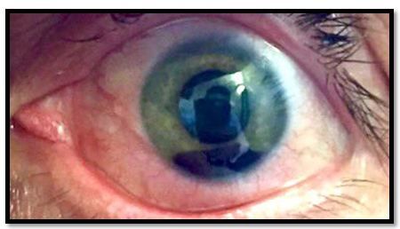
Figure 1. Facial image (please click to enlarge)

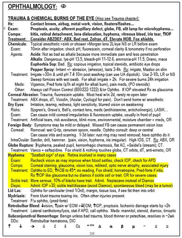
Figure 2. Page shot (please click to enlarge)

History: A 40-year-old African American man presents to the ED for an eye injury. He was playing racquetball without eye protection and made the mistake of looking back as his friend was hitting the ball. The ball struck him directly in his left eye without first bouncing off a wall. He complains of pain and severe vision loss but denies double vision, loss of consciousness, or other complaints.
Exam: There is an obvious hyphema that takes up about 33% of the anterior chamber of the left eye (image not available). You also note a dilated and slightly irregular pupil. Vision is hand motion only in the affected eye.
The image seen in Figure 1 (please click to enlarge) is from the day after the injury when the hyphema had settled fully and therefore appears smaller.
Initial impression: Traumatic hyphema
Questions
1. What testing should be done for this patient?
2. What is the usual treatment for hyphema?
3. Are there any medical conditions that change treatment?
Answers and discussion on next page
Answers
1. What testing should be done for this patient? Measure the intraocular pressure (IOP) and do a sickle prep.
2. What is the usual treatment for hyphema? See page shot below.
3. Are there any medical conditions that change treatment? Yes, sickle disease or trait.
Discussion
Hyphema usually presents with pain and/or blurry vision after trauma to the eye. Any patient who suffers eye or facial trauma should be checked for hyphema. Early on, blood will be diffusely floating in the anterior chamber and will gradually settle with gravity. Vision may improve as this happens and the hyphema may change size and density. It is important to be aware that other, less visible, eye injuries may also be present so an ophthalmologist should see the patient in the ED for a thorough eye exam.
There are many other potential complications requiring specialty follow up (see Page Shot in Figure 2, above, for more information--please click on image to enlarge).
Treatment for hyphema involves rest with the head elevated at least 45 degrees as well as topical agents (more information in Page Shot in Figure 2). If IOP is elevated the usual medications for acute glaucoma should be administered with the caveat that Diamox should be avoided if the patient has sickle disease or sickle trait. African Americans should therefore be screened for sickle trait as there is an approximately 10% rate of asymptomatic carriers. Patients with elevated IOP or sickle cell disease should be admitted.
Case Conclusion: IOP was >30. Patient was admitted overnight for aggressive medical treatment and bed rest. Anterior chamber washout in the OR was considered but the patient improved significantly without it.
For additional information on diagnosis and treatment of eye trauma, including hyphema, please see The Emergency Medicine 1-Minute Consult Pocketbook.
