- Clinical Technology
- Adult Immunization
- Hepatology
- Pediatric Immunization
- Screening
- Psychiatry
- Allergy
- Women's Health
- Cardiology
- Pediatrics
- Dermatology
- Endocrinology
- Pain Management
- Gastroenterology
- Infectious Disease
- Obesity Medicine
- Rheumatology
- Nephrology
- Neurology
- Pulmonology
Ocular Signs of Genetic Disease
Photo quiz

Case 1:
A 21-year-old man seeks medical attention because of weight loss, unclear speech, and diminished athletic performance. He has noted progressive difficulty in filling out forms that require identification numbers, occasional difficulty in swallowing or pronouncing certain syllables, and an increase in his golf scores from scratch to the mid-80s.
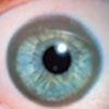
The examination reveals normal vision, intermittent oral dystonia with garbled speech, and a pronounced tremor with arm extension. His irides appear as shown. A complete blood cell count and renal function tests are normal; however, there is a 3-fold increase in serum aspartate aminotransferase and alanine aminotransferase levels.
What is the most likely diagnosis-and what further testing would you consider?
(Answer on next page.)

Case 1: Wilson disease
The photograph of this man's iris shows the Kayser-Fleischer ring that is characteristic of Wilson disease, an autosomal-recessive disorder. The patient's tremor, dysphagia, dysarthria, and deteriorating motor performance are typical manifestations of this disease. Neurological symptoms can include dystonia, dementia, and movement disorders resembling Parkinson disease. Other associated disorders include hepatic disease that can progress to severe cirrhosis and coma; renal disease that can include tubular dysfunction with aminoaciduria and/or calculi; hemolytic anemia; and skeletal disease with osteoporosis, osteomalacia, arthritis, and joint hypermobility. Symptoms can appear from adolescence to middle age; hepatic disease predominates in juvenile forms.1
Like the other disease manifestations, the deep-green or copper-colored (Kayser-Fleischer) ring on the periphery of the iris is caused by copper deposition. Confirmatory test results include a high serum copper level not bound to ceruloplasmin, and low ceruloplasmin and high urinary copper levels. Elevated copper levels reflect mutations in a gene responsible for copper transport-the adenosine triphosphatase, copper-transporting, β-polypeptide gene (ATP7B).
Recognition of Wilson disease is important because the hepatic and neurological manifestations can be treated with D-penicillamine, trientine, or zinc acetate. The incidence of Wilson disease is about 1 in 50,000 in the United States and somewhat higher worldwide.
AN ONLINE RESOURCE ON GENETIC DISORDERS
An extremely useful and convenient resource for information on genetic disorders is Online Mendelian Inheritance in Man (OMIM) (www.ncbi.nlm.nih.gov/sites/entrez?db=OMIM), an online database derived from the pioneering catalogue of Mendelian diseases compiled by Dr Victor McKusick. Entry of OMIM in your browser takes you to the search page, where entry of disease names or symptoms will yield the appropriate entries. McKusick assigned a unique number to each Mendelian or single gene disorder and started with 1 for autosomal-dominant, 2 for autosomal-recessive, 3 for X-linked, 4 for Y-linked, and 5 for mitochondrial diseases. Newer entries all start with a 6 that does not reflect the mechanism of inheritance. Another caveat about the OMIM catalogue is that some multifactorial or chromosomal disorders are listed, in part to record progress in finding single gene markers that are predictive of disease susceptibility (eg, diabetes mellitus or schizophrenia) or are located within key chromosome regions (eg, the RB retinoblastoma gene that was found through study of a chromosome 13 deletion).
Entry of "Wilson disease" on the OMIM search page yields entry #277900 that summarizes the history, clinical findings, and relevant biochemical/molecular data when available. Key references are provided, as well as links to PubMed for relevant abstracts, to www.genetests.org to see which laboratories offer DNA testing for the condition, and even to chromosome location or DNA sequence data available in the human genome database.
REFERENCE:
1. Taly AB, Meenakshi-Sundaram S, Sinha S, et al. Wilson disease: description of 282 patients evaluated over 3 decades. Medicine. 2007;86:112-121.

Case 2:
A 23-year-old woman is brought to the emergency department after being found unconscious in her apartment. She has a contusion on her head that is consistent with a fall. The custodian who found her said that she made some jerking movements before she became still and unresponsive.
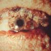
Which clues in the photograph (from a previous eye examination) suggest an underlying disorder? What steps would you take to determine the cause of the coma?
(Answer on next page.)

Case 2: Neurofibromatosis-1
The dark-brown Lisch nodules in the iris and small neurofibromata in the adnexa surrounding the eye are typical of neurofibromatosis-1, or NF-1 (OMIM #166200). Other associated findings are multiple caf-au-lait spots with smooth (coast of California) borders that distinguish them from less specific coffee-colored spots with ragged (coast of Maine) borders seen in disorders such as McCune-Albright syndrome (OMIM #174800). A survey in Michigan proposed a diagnostic criterion of 6 caf-au-lait spots greater than 1.5 cm; this criterion applies only to adults because the spots develop during the first 4 years of life, arising as purplish, frequently pruritic blotches.1 Axillary or inguinal freckling and skeletal complications, such as scoliosis or pseudoarthrosis, can also occur.
Although as many as 70% of persons with NF-1 have only caf-au-lait spots, complications can include benign tumors (schwannomas) that are malignant by position, leading to increased intracranial pressure and seizures, non-resectable plexiform neurofibromas, or renal artery compression with hypertension (pheochromocytomas). Some 10% of affected persons have mild mental disability, and there is a 2-fold increased risk of malignant tumors (neurofibrosarcoma, rhabdomyosarcoma, and many others). In this patient, a seizure was the most likely cause of her coma; a head MRI scan would be indicated to look for a tumor and/or for hydrocephalus.
NF-1 is an autosomal-dominant disease; the incidence is about 1 in 3000. The causative gene is known (neurofibromin). Gene testing is available but is not highly sensitive because of the large gene size and the variability of mutations.
Recognition of NF-1 is important for preventive health care, which includes monitoring for nerve compression, hypertension, and skeletal changes. Some experts recommend a baseline head MRI scan, which can show "unidentified bright objects" in the cerebellum and basal ganglia that are additional diagnostic criteria in children-these lesions do not increase the risk of tumors.2 Ophthalmological examination to demonstrate the Lisch spots (often visible only by slit-lamp) and deepening of the optic cup can be useful diagnostically and to monitor for another low-risk complication, glaucoma.
REFERENCES:
1. Crowe FW, Schull WJ, Neel JV. A Clinical, Pathological and Genetic Study of Multiple Neurofibromatosis. Springfield, Ill: Charles C. Thomas; 1956.
2. DeBella K, Poskitt K, Szudek J, Friedman JM. Use of "unidentified bright objects" on MRI for diagnosis of neurofibromatosis 1 in children. Neurology. 2000;54:1646-1651.

Case 3:
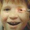
This 7-year-old boy is being evaluated for hereditary deafness. His left eye is brown, and his right eye is blue. He has been healthy except for sensorineural hearing loss that is not severe enough to require hearing aids or cochlear implants.
His mother and grandmother have complete hearing loss, and his mother has a forelock of white hair. His grandmother states that 2 of her children are deaf and 2 have normal hearing. The boy's sister has normal hearing.
Are this child's dissimilar irides related to the hereditary deafness in his family? Should this ocular finding prompt a search for other anomalies?
(Answer on next page.)

Case 3: Waardenburg syndrome
This boy has heterochromia with differently colored irides, a finding that can be isolated or can occur as part of the Waardenburg syndrome (OMIM #193500). The singer David Bowie and others with isolated heterochromia do not have neurosensory deficits.
Waardenburg syndrome is an autosomal-dominant disorder that consists of neurosensory deafness; widely spaced eyes (hypertelorism); and pigmentary changes that can include a white forelock (poliosis), premature graying, white patches on the skin (usually ventral midline), and heterochromia.1 The findings vary markedly even within the same family, as illustrated by more severe deafness in the boy's mother and grandmother. Occasional findings include cleft lip/palate, Sprengel deformity, vertebral anomalies, absent vagina, and uterine anomalies.
This broader spectrum of malformations might be anticipated because Waardenburg syndrome is caused by the PAX3 gene that shares considerable DNA sequence homology with the paired developmental gene of the fruit fly. A large fraction (30% to 50%) of Drosophila genes are conserved in mammals and have been frequently associated with human syndromes (eg, homeotic, or HOX, genes with human limb defects). Other types of Waardenburg syndrome with specific associations, such as Hirschsprung disease, have been defined. Although rare, Waardenburg syndromes are important to recognize in order to detect hearing loss and associated malformations. *
Author's note: The photographs in cases 1 and 2 are from Dr Harold Falls, an ophthalmologist who helped establish one of the first human genetics clinics, at the University of Michigan in 1941. He was one of the first to recognize X-linked inheritance in humans, exemplified by the ocular albinism that he described in 1951 as the Nettleship-Falls syndrome (OMIM #300100). Dr Falls attended clinical genetics rounds even in retirement and would quietly remind his laboratory-oriented colleagues that eyes can be the essence of diagnosis. He died recently at the age of 96 (www.umich.edu/~urecord/ 0506/Jun12_06/obits.shtml).
REFERENCE:
1. Hornyak TJ. The developmental biology of melanocytes and its application to understanding human congenital disorders of pigmentation. Adv Dermatol. 2006;22:201-218.
FOR MORE INFORMATION:
Wilson GN. Clinical Genetics: A Short Course. New York: John Wiley & Sons, Inc; 2000.
