- Clinical Technology
- Adult Immunization
- Hepatology
- Pediatric Immunization
- Screening
- Psychiatry
- Allergy
- Women's Health
- Cardiology
- Pediatrics
- Dermatology
- Endocrinology
- Pain Management
- Gastroenterology
- Infectious Disease
- Obesity Medicine
- Rheumatology
- Nephrology
- Neurology
- Pulmonology
Monitoring the response to therapy for pulmonary arterial hypertension, part 2
Despite the recent development of several new therapies, pulmonary arterial hypertension (PAH) remains an incurable disease. Careful monitoring of disease progression is vital to ensuring that patients receive maximal medical therapy before the onset of overt right-sided heart failure. In part 1 of this article, I reviewed the role of the history and physical examination, chest radiography, electrocardiography, echocardiography, and pulmonary artery catheterization. In part 2, I focus on MRI, cardiopulmonary exercise testing (CPET), the 6-minute walk test, and biomarkers.
Despite the recent development of several new therapies, pulmonary arterial hypertension (PAH) remains an incurable disease. Careful monitoring of disease progression is vital to ensuring that patients receive maximal medical therapy before the onset of overt right-sided heart failure. In part 1 of this article, I reviewed the role of the history and physical examination, chest radiography, electrocardiography, echocardiography, and pulmonary artery catheterization. In part 2, I focus on MRI, cardiopulmonary exercise testing (CPET), the 6-minute walk test, and biomarkers.
MRI
This diagnostic test combines several advantages of pulmonary artery catheterization and transthoracic echocardiography (TTE). Like TTE, MRI is noninvasive and can be done repeatedly with little harm to the patient. At the same time, it can provide most of the hemodynamic measurements obtained by catheterization. Its ability to more precisely visualize right ventricular (RV) structures allows accurate measurements of RV end-diastolic and end-systolic dimensions, and computer processing of multiple cross-sectional areas provides ventricular volume and mass.
The RV ejection fraction can be calculated as the difference between RV end-diastolic volume and RV end-systolic volume. The RV stroke volume can be measured by examining volumetric flow in the pulmonary artery, and RV output can be calculated from the product of RV stroke volume and heart rate. Maximal systolic cross-sectional area and mean systolic blood flow velocity over the main pulmonary artery can be used to calculate the mean pulmonary artery pressure (PAP). Studies of patients with PAH have shown good agreement between mean PAP measured by MRI and by pulmonary artery catheterization.1
MRI can also assess RV function by generating pressure-volume loops from measurements of ventricular volumes and pressures.2 RV myocardial contractility can be assessed by the slope of the end-systolic pressure-volume relationship, a load-independent parameter of myocardial contractility.
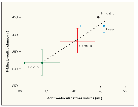
MRI has been used to monitor improvement in RV function in numerous clinical trials in patients with PAH. Roeleveld and colleagues3 found increased RV mass, RV end-diastolic volume, and RV end-systolic volume at baseline in 9 patients with PAH. After treatment with epoprostenol, RV end-diastolic volume was unchanged, but RV stroke volume and functional capacity improved (Figure 1). Repeated right heart catheterization revealed no change in mean PAP but a significant increase in cardiac output, suggesting that epoprostenol improved performance by increasing RV contractility and reducing pulmonary vascular resistance (PVR). These findings suggest that MRI may be a useful tool for monitoring RV functional responses to therapy in patients with PAH.

Figure 1 – The 6-minute walking distance improves as the right ventricular stroke volume increases in patients with pulmonary arterial hypertension who were treated for 12 months with epoprostenol. (Adapted from Roeleveld RJ et al. Chest. 2004.3)
Cardiopulmonary exercise testing
CPET is a noninvasive way of assessing cardiopulmonary performance during exercise. Patients ride a stationary bike or walk a treadmill while breathing through an airtight mouthpiece that continually measures the Po2 and Pco2 of exhaled air. By measuring the volume of gas inhaled and exhaled per unit of time, oxygen consumption (Vo2) and carbon dioxide elimination (Vco2) can be measured in mL/min. Cardiac output can be determined from Vo2 based on the Fick equation:
Q = Vo2/[C(a-v)o2]
The arteriovenous oxygen difference-C(a-v)o2-cannot be determined precisely without measurement of mixed venous oxygen saturation. However, at maximal exercise, C(a-v)o2 is about 75% of total oxygen carrying capacity of the blood, which can be measured if the patient’s arterial oxygenation saturation and hemoglobin level are known. Vo2 is determined by cardiac output, pulmonary gas exchange, and the extraction and utilization of oxygen by skeletal muscle.
Patients with PAH may have impaired RV output as a result of elevated PVR as well as reduced gas exchange as a result of pulmonary vascular disease, which leads to pruning of the pulmonary vascular bed. Also, oxygen uptake and utilization are often impaired by deconditioning. Thus, an abnormal exercise test result is not uncommon in patients with PAH, and it does not indicate which part of the respiratory cycle is compromised.
However, CPET can provide important information about the overall health of the patient. In the non- deconditioned patient, a rise in maximal Vo2 strongly suggests improvement in pulmonary vascular disease and is a more objective measurement of cardiopulmonary function than a submaximal exercise test. As a result, maximal Vo2 has been used as a primary outcome measure in clinical studies of PAH.4-6 However, some studies have shown no increase in maximal Vo2 despite an increase in 6-minute walk distance.
Another way of assessing cardiac output is to divide Vo2 by the pulse rate. Considering Q = Vo2/[C(a-v)o2] and Q = SV × HR (SV is stroke volume and HR is heart rate), SV × HR = Vo2/[C(a-v)o2] or SV × [C(a-v)o2] = Vo2/HR.
Assuming that the arteriovenous oxygen difference remains constant for a given work load, stroke volume should be directly proportional to Vo2/HR. This measurement is referred to as the oxygen pulse and is used as a noninvasive method for assessing the amount of oxygen extracted per heartbeat. Normally, the oxygen pulse increases rapidly with exercise and becomes asymptotic toward maximal exercise.7 A low, flat oxygen pulse with increasing work suggests reduced stroke volume, but it can also occur from failure of the skeletal muscle to increase oxygen extraction.
Thus, a low oxygen pulse can be seen with deconditioning, cardiovascular disease, and early exercise limitation resulting from ventilatory constraint. However, in any given individual, an increase in oxygen pulse in response to therapy suggests improving cardiopulmonary performance and, as such, can be used to monitor response to therapy in patients with PAH.
In clinical studies, patients with PAH have been found to have lower mean Vo2, oxygen pulse, and anaerobic threshold and increased minute ventilation/Vco2 than controls.8,9 Furthermore, the mean Vo2 and oxygen pulse have been demonstrated to correlate closely with 6-minute walking distance and with improvement in the response to pulmonary vasodilator therapy.6,10,11
6-Minute walk test
Maximal exercise tests offer valuable information regarding peak cardiopulmonary function, but they are not suitable for use in patients with significant functional impairment. The 6-minute walk test is a submaximal stress test that was developed for monitoring exercise performance in patients with chronic heart disease to help determine their capacity to undertake day-to-day activities.12
During this test, the patient walks unassisted on a horizontal surface at a comfortable, but steady pace for 6 minutes. Patients can stop to rest as often as they feel is necessary but are instructed to continue walking as soon as they can. Guidelines on how the test should be performed have been published to ensure uniformity between testing centers and suggest that patients walk at least 100 ft in a straight line before turning around.13 Blood pressure, pulse rate, and arterial oxygen saturation are measured at the beginning and end of the test. At the conclusion of the walk, patients are asked to rate their dyspnea on a scale of 1 to 10 (Borg dyspnea score).
Miyamoto and associates10 found that the average 6-minute walking distance in patients with PAH is less than half that of healthy age- and sex-matched controls (297 m ± 188 m versus 655 m ± 91 m; P < .001). The 6-minute walking distance correlated modestly with baseline cardiac output and total pulmonary resistance, although not with PAP. A 6-minute walking distance greater than 332 m was the only noninvasive measure of pulmonary hypertension that independently predicted survival over a 2-year period. Similarly, Paciocco and associates14 found that a 6-minute walk distance of 300 m or less was associated with a 2.4-fold increase in mortality risk and a fall in oxygen saturation of 10% or more was associated with a 2.9-fold increase in mortality risk.
The 6-minute walking distance has become the standard exercise test used to assess functional response for virtually every medical therapy trial. Most trials report an average placebo-corrected increase in 6-minute walking distance of about 30 to 50 m after 3 months of therapy.15-19
In general, the patient’s 6-minute walking distance should be monitored every 4 to 6 months in patients receiving therapy for PAH, or whenever the clinician suspects that functional status is declining. In addition, most patients with PAH should perform moderate exercise regularly. We recommend patients walk for at least 6 minutes on level ground at home several times a week.
Biomarkers
Acute and chronic RV failure has been associated with increased circulating levels of plasma brain natriuretic peptide (BNP) and troponin. However, neither of these proteins is specific for the right ventricle, and the possibility of left ventricular disease or strain needs to be considered in evaluating changes in either marker. Elevations in levels of BNP and troponin are considerably smaller in patients with right-sided heart failure than in patients with left-sided heart disease. Plasma BNP levels above 200 pg/mL or troponin levels greater than 0.15 ng/mL are not usually considered to be pathognomonic of cardiac disease, but they can indicate significant RV dysfunction in patients with PAH.
Atrial natriuretic peptide and BNP are released by the cardiac atria and ventricles in response to several stimuli, including myocardial distention, tachycardia, and hypoxia. Experimental pulmonary hypertension in animals causes an increase in RV messenger RNA and protein levels and circulating levels of both peptides.20 Patients with PAH or chronic thromboembolic pulmonary hypertension have higher plasma BNP levels than patients with RV volume overload or age-matched controls.21 Furthermore, circulating BNP levels correlate directly with PAP, PVR, and right atrial pressure and are inversely related to cardiac index, 6-minute walk distance, and peak oxygen uptake.22
An elevated BNP level at presentation indicates a worse prognosis in patients with PAH, as does failure to reduce plasma BNP levels during treatment. Nagaya and colleagues23 found that a plasma BNP level above 150 pg/mL at presentation and failure to decrease plasma BNP levels below 180 pg/mL after 3 ± 1 month of therapy were associated with greater 2-year mortality. Similar findings have been reported with N-terminal pro-BNP levels greater than 1400 pg/mL and with higher plasma endothelin-1 levels (Figure 2).24,25 Some studies suggest that N-terminal pro-BNP, the precursor of BNP, may be a more sensitive measure of RV strain than BNP.26
Figure 2 – The relationship between brain natriuretic peptide (BNP) and endothelin-1 (ET-1) levels and survival or need for transplant are shown for 88 patients with pulmonary arterial hypertension. Data shown are divided by quartiles. The highest BNP quartile (plasma BNP level > 443 pg/mL; P = .029) and ET-1 quartile (ET-1 level > 17.3 pg/mL; P = .001) were associated with worse outcome. (Adapted from Frantz RP et al. Am J Respir Crit Care Med. 2008.25)
Cardiac troponin is a sensitive indicator of myocardial ischemia, and increases in circulating levels are common in clinical situations in which myocardial oxygen demand exceeds supply without acute obstruction to coronary blood flow. The rise in RV pressure in patients with progressive PAH increases RV wall tension and oxygen demand. Torbicki and associates27 found elevated troponin levels in 14% of patients with PAH or inoperable chronic thromboembolic pulmonary hypertension. The 6-minute walk distance and mixed venous oxygen saturation were lower in patients with elevated troponin levels, and cumulative 24-month survival was only 29%, compared with 81% in patients with normal troponin levels.

Summary
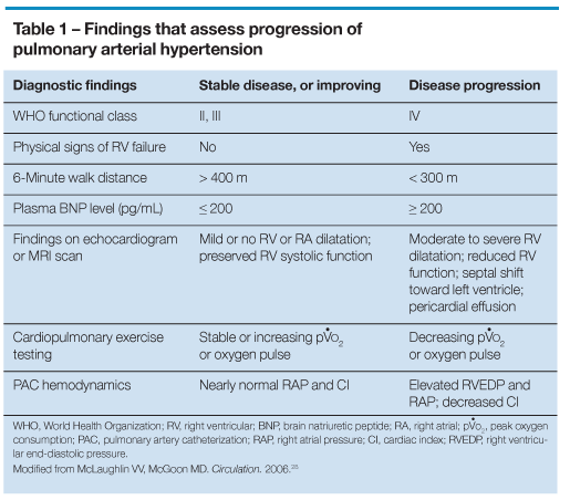
Using the techniques that we have described in conjunction with a careful history and physical examination, the practitioner can make a reasonable assessment of PVR and right-sided heart performance. Comparison of serial measurements usually provides a clear picture of the patient’s response to therapy, but no one technique provides all the necessary information. Maintaining a chart of test results for each patient frequently helps clarify who is improving and who is getting worse (Table 1).28
When the results of most monitoring tools point in the same direction, a patient’s prognosis is fairly clear and treatment can be adjusted as indicated. However, it is not uncommon for patients to have evidence of disease progression on some tests and signs of improvement on others. Under these circumstances, pulmonary artery catheterization may help accurately assess cardiopulmonary performance.

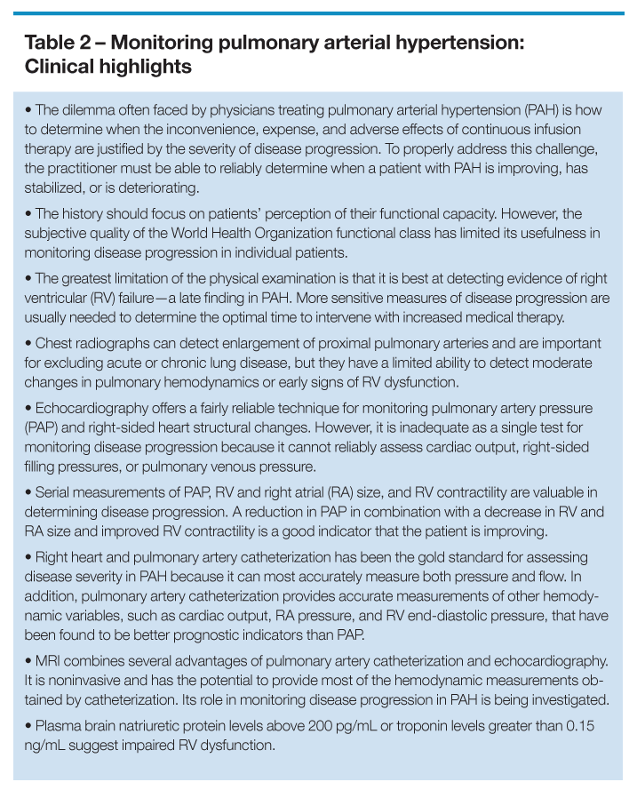
Another generalization may be worth keeping in mind: Patients with PAH who are “straddling the fence” between better and worse after several months of therapy are more likely to decline than to slowly improve. When it doubt, it is better to assume that a patient’s condition is worsening until repeated testing demonstrates otherwise. Table 2 presents clinical highlights of the monitoring process.
References:
REFERENCES
1. Laffon E, Vallet C, Bernard V, et al. A computed method for noninvasive MRI assessment of pulmonary arterial hypertension. J Appl Physiol. 2004;96:463-468.
2. Kuehne T, Yilmaz S, Steendijk P, et al. Magnetic resonance imaging analysis of right ventricular pressure-volume loops: in vivo validation and clinical application in patients with pulmonary hypertension. Circulation. 2004;110:2010-2016.
3. Roeleveld RJ, Vonk-Noordegraaf A, Marcus JT, et al. Effects of epoprostenol on right ventricular hypertrophy and dilatation in pulmonary hypertension. Chest. 2004;125:572-579.
4. Barst RJ, McGoon M, McLaughlin V, et al; Beraprost Study Group. Beraprost therapy for pulmonary arterial hypertension [published correction appears in J Am Coll Cardiol. 2003;42:591]. J Am Coll Cardiol. 2003;41:2119-2125.
5. Hoeper MM, Taha N, Bekjarova A, et al. Bosentan treatment in patients with primary pulmonary hypertension receiving nonparenteral prostanoids. Eur Respir J. 2003;22:330-334.
6. Wax D, Garofano R, Barst RJ. Effects of long-term infusion of prostacyclin on exercise performance in patients with primary pulmonary hypertension. Chest. 1999;116:914-920.
7. American Thoracic Society, American College of Chest Physicians. ATS/ACCP statement on cardiopulmonary exercise testing. Am J Respir Crit Care Med. 2003;167:211-277.
8. D’Alonzo GE, Gianotti LA, Pohil RL, et al. Comparison of progressive exercise performance of normal subjects and patients with primary pulmonary hypertension. Chest. 1987;92:57-62.
9. Deboeck G, Niset G, Lamotte M, et al. Exercise testing in pulmonary arterial hypertension and in chronic heart failure. Eur Respir J. 2004;23:747-751.
10. Miyamoto S, Nagaya N, Satoh T, et al. Clinical correlates and prognostic significance of six-minute walk test in patients with primary pulmonary hypertension. Comparison with cardiopulmonary exercise testing. Am J Respir Crit Care Med. 2000;161(2 pt 1):487-492.
11. D’Alonzo GE, Gianotti L, Dantzker DR. Noninvasive assessment of hemodynamic improvement during chronic vasodilator therapy in obliterative pulmonary hypertension. Am Rev Respir Dis. 1986;133:380-384.
12. Guyatt GH, Sullivan MJ, Thompson PJ, et al. The 6-minute walk: a new measure of exercise capacity in patients with chronic heart failure. Can Med Assoc J. 1985;132:919-923.
13. ATS Committee on Proficiency Standards for Clinical Pulmonary Function Laboratories. ATS statement: guidelines for the six-minute walk test. Am J Respir Crit Care Med. 2002;166:111-117.
14. Paciocco G, Martinez FJ, Bossone E, et al. Oxygen desaturation on the six-minute walk test and mortality in untreated primary pulmonary hypertension. Eur Respir J. 2001;17:647-652.
15. Barst RJ, Rubin LJ, Long WA, et al. A comparison of continuous intravenous epoprostenol (prostacyclin) with conventional therapy for primary pulmonary hypertension. The Primary Pulmonary Hypertension Study Group. N Engl J Med. 1996;334:296-302.
16. Rubin LJ, Badesch DB, Barst RJ, et al. Bosentan therapy for pulmonary arterial hypertension [published correction appears in N Engl J Med. 2002;346:1258]. N Engl J Med. 2002;346:896-903.
17. McLaughlin VV, Gaine SP, Barst RJ, et al; Treprostinil Study Group. Efficacy and safety of treprostinil: an epoprostenol analog for primary pulmonary hypertension. J Cardiovasc Pharmacol. 2003;41:293-299.
18. Galiè N, Ghofrani HA, Torbicki A, et al; Sildenafil Use in Pulmonary Arterial Hypertension (SUPER) Study Group. Sildenafil citrate therapy for pulmonary arterial hypertension [published correction appears in N Engl J Med. 2006;354:2400-2401]. N Engl J Med. 2005;353:2148-2157.
19. Galiè N, Olschewski H, Oudiz RJ, et al. Ambrisentan in Pulmonary Arterial Hypertension, Randomized, Double-Blind, Placebo-Controlled, Multicenter, Efficacy Studies (ARIES) Group. Ambrisentan for the treatment of pulmonary arterial hypertension: results of the ambrisentan in pulmonary arterial hypertension, randomized, double-blind, placebo-controlled, multicenter, efficacy (ARIES) study 1 and 2. Circulation. 2008;117:3010-3019.
20. Hill NS, Klinger JR, Warburton RR, et al. Brain natriuretic peptide: possible role in the modulation of hypoxic pulmonary hypertension. Am J Physiol. 1994;266(3 pt 1):L308-L315.
21. Nagaya N, Nishikimi T, Okano Y, et al. Plasma brain natriuretic peptide levels increase in proportion to the extent of right ventricular dysfunction in pulmonary hypertension. J Am Coll Cardiol. 1998;31:202-208.
22. Leuchte HH, Holzapfel M, Baumgartner RA, et al. Clinical significance of brain natriuretic peptide in primary pulmonary hypertension. J Am Coll Cardiol. 2004;43:764-770.
23. Nagaya N, Nishikimi T, Uematsu M, et al. Plasma brain natriuretic peptide as a prognostic indicator in patients with primary pulmonary hypertension. Circulation. 2000;102:865-870.
24. Fijalkowska A, Kurzyna M, Torbicki A, et al. Serum N-terminal brain natriuretic peptide as a prognostic parameter in patients with pulmonary hypertension. Chest. 2006;129:1313-1321.
25. Frantz RP, Robbins IM, Durst LA, et al. Endothelin-1 and BNP plasma levels predict survival in patients with pulmonary arterial hypertension. Am J Respir Crit Care Med. 2008;177:A535.
26. Leuchte HH, El Nounou M, Tuerpe JC, et al. N-terminal pro-brain natriuretic peptide and renal insufficiency as predictors of mortality in pulmonary hypertension. Chest. 2007;131:402-409.
27. Torbicki A, Kurzyna M, Kuca P, et al. Detectable serum cardiac troponin T as a marker of poor prognosis among patients with chronic precapillary pulmonary hypertension. Circulation. 2003;108:844-848.
28. McLaughlin VV, McGoon MD. Pulmonary arterial hypertension. Circulation. 2006;114:1417-1431.
