- Clinical Technology
- Adult Immunization
- Hepatology
- Pediatric Immunization
- Screening
- Psychiatry
- Allergy
- Women's Health
- Cardiology
- Pediatrics
- Dermatology
- Endocrinology
- Pain Management
- Gastroenterology
- Infectious Disease
- Obesity Medicine
- Rheumatology
- Nephrology
- Neurology
- Pulmonology
A Middle-Aged Man With Recurrent Pneumonia and Renal Failure
A 56-year-old was seen in the ED after 4 days of hemoptysis and intermittent left chest pain. He also complained of exertional dyspnea and arthralgias. He had been treated for “pneumonia” twice during the past month. Histories were unremarkable.
A 56-year-old man presented to the ED after 4 days of hemoptysis and intermittent left chest pain that worsened when he was supine. Ibuprofen provided some relief. He also felt tired and complained of exertional dyspnea. Joint pain, primarily affecting the knees, shoulders, and elbows also had bothered him for the past few months. His temperature was normal; he had no chills and had not lost any weight. He said he had been treated for “pneumonia” twice during the past month. Past medical and surgical histories were unremarkable. He was a social drinker and did not smoke cigarettes.
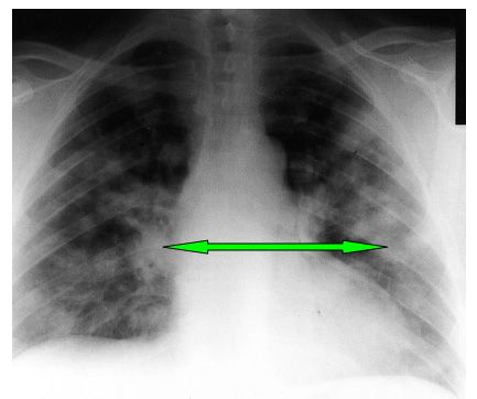
Figure 1.
Arrows indicate bilateral infiltrates.
The patient was pale and had small telangiectasias on his face. He had mild tenderness over the knees and shoulders. Active and passive range of motion measures were normal. Results of blood chemistry tests were: sodium, 135 mEq/L; potassium, 5.3 mEq/L; chloride, 107 mEq/L; bicarbonate, 15 mEq/L; BUN, 91 mg/dL; creatinine, 7.9 mg/dL; glucose, 81 mg/dL. The hemoglobin level was 8.4 g/dL; white blood cell (WBC) count, 10.9 x 10³/μL; platelets, 234 x 103/μL. Urinalysis revealed large amounts of blood, and 82 red blood cells (RBCs) per high-power field. Chest films revealed bilateral infiltrates (Figure 1).
What’s Your Diagnosis? Answer on Next Page…
Diagnosis: Wegener granulomatosis.
After serologic investigation was positive for C-ANCA (cytoplasmic-antineutrophil cytoplasmic antibodies) directed against proteinase-3 (PR3) a renal biopsy was performed. Biopsy results showed pauci-immune necrotizing glomerulonephritis with cellular crescent formation (Figure 2a), consistent with Wegener granulomatosis (WG). Figure 2b shows normal renal histology.
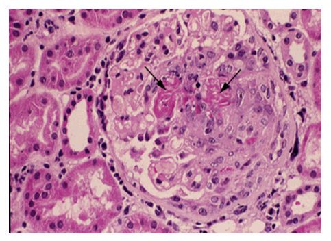
Figure 2a.
Arrows mark necrotizing lesions with red fibrin deposition.
The patient had received 2 rounds of dialysis and pulse corticosteroids before diagnosis. After the diagnosis was made, he was transferred to a tertiary medical center for definitive treatment with plasmapheresis, corticosteroids, cyclophosphamide, and dialysis. His symptoms have improved although his renal function remains unchanged since admission.
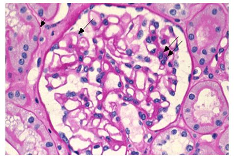
Figure 2b.
Arrows indicate normal glomeruli within the kidney.
Discussion
WG is a rare small-vessel vasculitis caused by inflammatory and granulomatous infiltrates. The disease affects persons of any age, but the median is 50 years. Men and women are affected equally. Although it is a multi-organ disease, WG most commonly affects the respiratory system and kidneys. Initial symptoms are vague (eg, fever, malaise, weight loss, and arthralgias). The upper respiratory system is involved in 95% of patients. Patients often have sinus tenderness, and purulent or bloody rhinorrhea. Saddle nose deformity is a characteristic result of nasal septal perforation (Figure 3).
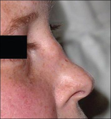
Figure 3.
Saddle nose deformity.
Photo courtesy of Raja Shekhar R Sappatibiyyani, MD
The upper airway lesions in the sinuses and nose classically show inflammation, necrosis, and granuloma formation. Chest radiographs reveal patchy or diffuse infiltrates, commonly mistaken for signs of bacterial pneumonia. Cavitary lesions also occur, raising suspicion for carcinomas. Common clinical complaints are cough, dyspnea, hemoptysis, and pleuritic chest pain. Renal disease is present in about three-quarters of patients. Urinalysis shows proteinuria, hematuria, and red cell casts. BUN and creatinine levels are typically elevated.
WG can also affect the eyes, ears, mouth, and skin. Ocular involvement can cause conjunctivitis, corneal ulcers, episcleritis, proptosis, or vision loss. Serous otitis media and hearing loss may follow eustachian tube inflammation. Oral ulcers are common and skin lesions appear as small blisters, ulcers, or nodules.
Routine laboratory tests are nonspecific for WG. However, characteristic findings include elevated ESR, anemia, elevated WBC and platelet counts, and mildly elevated rheumatoid factor.1,3,4
Diagnosis of WG is based on tissue biopsy of an involved organ, most commonly the kidney. Segmental, necrotizing glomerulonephritis with pauci-immune crescent formation is pathognomic. In approximately 90% of patients, serology is positive for C-ANCA, PR3.1,4,5
A positive C-ANCA result is 87% sensitive and 100% specific for WG, if checked while the disease is active.1,4 Immunoflourescence assay is most commonly used to measure serum C-ANCA. The resulting pattern shows heavy cytoplasmic staining and non-reactive nuclei (Figure 4).6
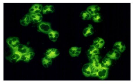
Figure 4.
Indirect immunofluorescence with heavy staining
in the cytoplasm; nuclei (clear areas) are nonreactive.
The American College of Rheumatology has established 4 criteria for the classification of WG,2 of which must be met for inclusion4:
• Abnormal urinary sediment (red cell casts or >5 RBC)
• Abnormal findings on CXR (cavities, infiltrates)
• Oral ulcers or nasal discharge
• Granulomatous inflammation on organ biopsy
Therapy depends on disease severity. Most regimens for local and early disease include methotrexate or cyclophosphamide. Other regimens include corticosteroids and azathioprine. The most severely ill patients often require dialysis and plasmapheresis, in addition to immunosuppressive therapy and corticosteroids.1-3 Half of these patients will have disease relapse within 2 years. Five-year survival rates approach 82% in patients treated with cyclophosphamide; however, 90% have disease-related morbidity.2
Teaching Points:
• In a patient with a long history of renal and pulmonary involvement, the differential diagnosis should include Wegener granulomatosis and Goodpasture syndrome
• Serology found positive for C-ANCA and pauci-immune crescent formation found on renal biopsy are highly diagnostic of Wegener granulomatosis
• The differential diagnosis for Wegener granulomatosis includes Churg-Strauss syndrome, microscopic polyangiitis, tumors of the upper airway or lung, chronic sinusitis, or sarcoidosis, as well as Goodpasture syndrome
References:
1. Langford CA, Fauci AS. The vasculitis syndromes. In: Kasper DL, Braunwald E, Fauci AS, et al, eds. Harrison’s Principles of Internal Medicine. 16th ed. New York: McGraw Hill Medical Publishing Division; 2005:2004-2006.
2. Haddow S, Brocato J. Wegener’s granulomatosis. Essential Evidence Plus. January 21, 2010. Available at: www.essentialevidenceplus.com.
3. Goldman J, Bennett B. Cecil Textbook of Medicine. 21st ed. Philadelphia: WB Saunders Company; 2000:1529-1532.
4. Klippel JH, Stone JH, Crofford LG, et al, eds. Primer on the Rheumatic Diseases. 13th ed. New York: Springer Science & Business Media, LLC; 2008:416-425.
5. Foster C, Mistry NF, Peddi PF, Sharma S, eds. The Washington Manual of Medical Therapeutics. 33rd ed. Philadelphia: Lippincott Williams and Wilkins; 2010.
6. Tappouni R, Burns A. Pituitary involvement in Wegener’s granulomatosis. Nephrol Dial Transplant. 2000;15:2057-2058.
7. Glassock R, Appel G. Clinical Spectrum of Antineutrophil Cytoplasmic Antibodies. Available at: www.uptodate.com/contents/clinical-spectrum-of-antineutrophil-cytoplasmic-antibodiesune. Accessed June 2011.
