- Clinical Technology
- Adult Immunization
- Hepatology
- Pediatric Immunization
- Screening
- Psychiatry
- Allergy
- Women's Health
- Cardiology
- Pediatrics
- Dermatology
- Endocrinology
- Pain Management
- Gastroenterology
- Infectious Disease
- Obesity Medicine
- Rheumatology
- Nephrology
- Neurology
- Pulmonology
Mediastinal Abscess From Laryngeal Mask Airway
The laryngeal mask airway (LMA) has become a popularalternative to endotracheal intubation. Many cliniciansconsider it a safe procedure, but complications do occur.Although uncommon, retropharyngeal perforation withmediastinal abscess can become a life-threatening event. Wereport a case of mediastinal abscess in an 84-year-old womanwho received LMA ventilation during a surgical procedurefor total knee replacement. [Infect Med. 2008;25:180-185]
The laryngeal mask airway (LMA) has been used in millions of patients with minimal complications since the 1980s. Reported complications include aspiration, local trauma during placement, and injuries caused by high cuff pressure and volume. A previous case of esophageal perforation, which was fatal, occurred as a complication of elective general anesthesia for cataract extraction in a 77- year-old woman.1 The patient had a difficult tracheal intubation, which occurred in the setting of a clinical trial of an intubating laryngeal mask. Besides this case, we have not found any previous report of retropharyngeal or mediastinal abscess secondary to LMA-associated retropharyngeal perforation.
Case report
Our patient was an otherwise healthy 84-year-old woman who underwent an elective left total knee replacement for degenerative osteoarthritis on November 4, 2005. She received spinal anesthesia and had spontaneous breathing with a No. 3 disposable LMA (LMA Unique, manufactured by LMA North America, Inc, San Diego). The LMA was inserted without the use of any instrument. She was anesthetized for 2 hours and 25 minutes. No complications were reported during anesthesia. No emesis or wretching occurred. The patient did not have a nasogastric tube. Subcutaneous emphysema of the neck and chest were noted in the recovery room.
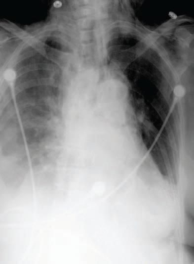
Postoperatively, the patient was stable, although she complained of a sore throat and hoarseness. The physical examination findings were remarkable for crepitations in the neck and supraclavicular fossae. On the first postoperative day (November 5), the chest radiograph revealed subcutaneous air in the neck and a pneumomediastinum that persisted the following day (Figure 1). The patient had a history of penicillin allergy. The empirical antibiotic coverage was switched from levofloxacin and metronidazole to cefotaxime and clindamycin.

Figure 1 - A chest radiographobtained November 5, 2005,shows subcutaneous emphysemain the left side of the neck andsuggestion of pneumomediastinum.The diameter of thesuperior mediastinum iswithin normal limits.Bilateral pulmonaryinfiltrates are greaterin the right lungthan in the left lung.

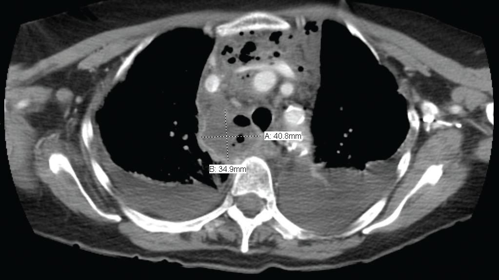
Figure 3 - A CT scan of the chest (horizontal view) obtained November 9, 2005, shows a fluiddensity structure, roughly measuring 3.5 & chi; 4.1 & chi; 6.3 cm, in the right posterior mediastinum.It is consistent with an abscess with extension into the anterior mediastinum between the superiorvena cava and the remainder of the central great vessels. Mass effect on the trachea is notedwith tracheal deviation to the left. Small bilateral pleural effusions are present with bibasilaratelectasis rather than consolidation.

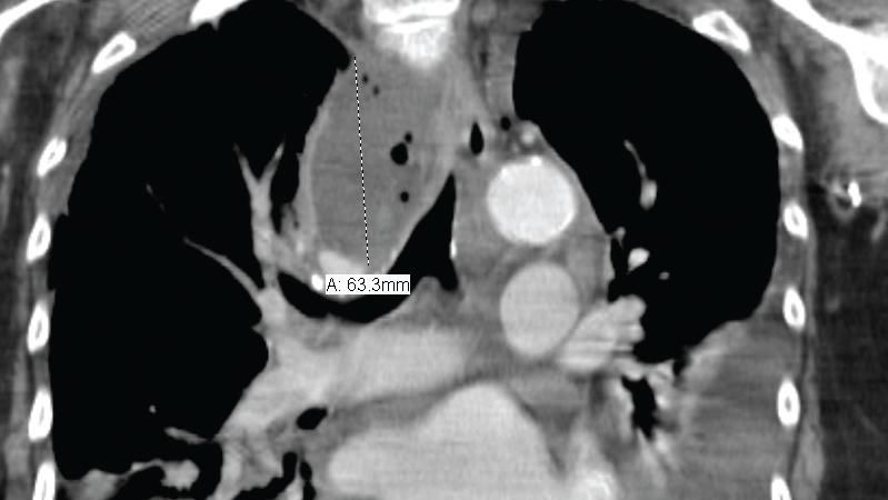
Figure 4 - ACT scan of the chest (coronal view) obtained November 9, 2005, shows extensivebilateral subcutaneous emphysema involving the base of the neck and extending into the deepcervical regions. Diffuse pneumomediastinum is also present without evidence of pneumothorax.Mild epicardial fat stranding is noted, consistent with inflammatory changes around thepericardium.
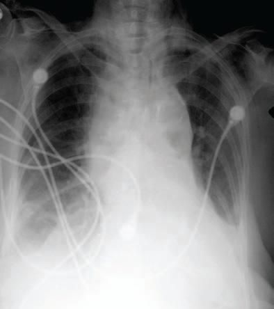
On November 7, the patient was noted to be more lethargic, but the vital signs remained stable and there was no evidence of respiratory distress. The patient was evaluated by thoracic surgery. A portable chest radiograph showed widening of the superior mediastinum with small bilateral pleural effusions (Figure 2).

Figure 2 - A chest radiographobtained November 7, 2005,shows that the transverse diameterof the superior mediastinumhas increased in comparisonto what was visualized inthe previous radiograph.
On November 8, the patient was alert and responsive. She denied any pains, sore throat, or difficulty in breathing. CT scans of the chest and neck obtained on November 9 documented evolving retropharyngeal and superior-posterior mediastinal abscesses (Figures 3 and 4).
On November 10, the patient underwent surgical drainage; approximately 100 mL of purulent material was drained from the retropharyngeal space and mediastinum viaright cervical exploration with open drainage of retropharyngeal and mediastinal abscesses.
The patient's preoperative white blood cell (WBC) count increased from 7100/& mu;L to 20,400/& mu;L, with 33% bands. After surgical drainage of the abscesses, the WBC count decreased to 11,000/& mu;L without bands.
The Gram stain of the abscesses was compatible with oral flora and included multiple Grampositive and Gram-negative organisms. Cultures of the purulent material obtained on November 10 showed coagulase- negative staphylococci, metronidazole- and clindamycin- resistant Lactobacillus acidophilus, Candida albicans, and clindamycin-resistant Prevotella melaninogenica. Fluconazole was added to the patient's antimicrobial regimen on November 11, and after bacteriological results were obtained, treatment with vancomycin and metronidazole was started. Thoracentesis performed on November 14 showed a sterile exudate.
The patient slowly improved without the need for further intervention after surgical drainage. She received 6 weeks of intravenous antibiotic therapy. The posterior pharyngeal tear healed, and a residual pocket in the prevertebral space closed spontaneously.
Discussion
A retropharyngeal tear with associated mediastinitis and abscess was a complication of LMA in our patient. She did not have any known predisposing factors or procedures that could have contributed to the incident. Such a complication related to LMA use has not been reported previously.
A retropharyngeal or mediastinal abscess that occurs as a complication of airway management requires rapid diagnosis, appropriate antibiotics, and surgical consultation. Recent reviews have made us aware of other potential complications. 2,3 The Agency for Healthcare Research and Quality previously reviewed some aspects of this case.4
Respiratory distress never developed in our patient. A lack of respiratory symptoms can lead to a false sense of security. Subcutaneous emphysema may be more common with positive pressure ventilation, which was not used in our patient. The earliest sign of retropharyngeal perforation was subcutaneous air. The patient had leukocytosis with left shift on the first postoperativeday, which suggested the presence of a deep tissue infection.
Radiological evidence of subcutaneous emphysema and pneumomediastinum appeared within 24 hours postoperatively. A widening mediastinum, detected radiographically, is an early sign of mediastinitis or abscess. Our patient showed a widened mediastinum and lethargy 3 days after surgery. ACT scan of the chest will confirm the presence of a potential retropharyngeal or mediastinal abscess. The patient did not improve until surgical drainage was performed and appropriate antibiotic therapy was begun.
LMA is generally a safe procedure. However, we must remain alert to the potential for known and unreported complications. The appearance of subcutaneous air during or after the procedure should prompt a rapid investigation as to its cause and possible complications.
References:
- Branthwaite MA. An unexpected complication of the intubating laryngeal mask. Anaesthesia. 1999;54:166-167.
- Divatia JV, Bhowmick K. Complications of endotracheal intubation and other airway management procedures. Indian J Anesth. 2005;49: 308-318.
- Pollack CV Jr. The laryngeal mask airway: a comprehensive review for the emergency physician. J Emerg Med. 2001;20:53-66.
- Secured But Not Always Safe. AHRQ Web M&M. November 2006. Available at: www. webmm.ahrq.gov/case.aspx?caseID=139. Accessed January 10, 2008.
