- Clinical Technology
- Adult Immunization
- Hepatology
- Pediatric Immunization
- Screening
- Psychiatry
- Allergy
- Women's Health
- Cardiology
- Pediatrics
- Dermatology
- Endocrinology
- Pain Management
- Gastroenterology
- Infectious Disease
- Obesity Medicine
- Rheumatology
- Nephrology
- Neurology
- Pulmonology
Man With Worsening Cough and Dyspnea
Man With Worsening Cough and Dyspnea
A previously healthy 49-year-old man who resides in the central valley area of California presents with a 4-week history of worsening dry, irritating cough and dyspnea. He can barely walk a few steps on level ground and has to sleep propped up.


At the onset of his illness, the patient had fever with rigors and profuse night sweats and felt exhausted. He was evaluated several times at different urgent care centers, and several courses of oral antibiotics were prescribed. These included azithromycin, trimethoprim/sulfamethoxazole, levofloxacin and, most recently, telithromycin. There was little improvement in his symptoms.
The patient has lost 16 lb in the past month and complains of poor appetite. He denies hemoptysis, chest pain, and ankle edema. He has no rash, nausea, vomiting, diarrhea, or abdominal pain. There is no history of headache, vision problems, weakness, paresthesias, syncope, seizures, urinary symptoms, or exposure to persons with tuberculosis or viral syndromes.
The patient is in a monogamous married relationship. He does not smoke, drink alcohol, or use illicit drugs. He has had a pet dog for the past 4 years. There is no history of transfusions, tattoos, or foreign travel.
The patient's mother has diabetes. His father has hypertension and coronary artery disease that are managed with oral medications.
Examination. This well-built, well-nourished man looks chronically ill and is in moderate respiratory distress. His heart rate is 94 beats per minute and regular; temperature, 37.3°C (99.1°F); respiration rate, 28 breaths per minute; and blood pressure, right upper limb, 132/72 mm Hg. Hydration status is good. Examination of the head and neck reveals no icterus, erythema, or evidence of candidal infection. There is no palpable adenopathy.
Respiratory examination reveals normal chest contours and symmetric movement. The trachea is centrally located. The chest is resonant to percussion. Harsh bronchovesicular breath sounds with prolonged expiration and diffuse bilateral wheezing are noted. The jugular vein pulse and the apex beat are normal. Heart sounds are normal with no murmur or gallop. Findings from the remainder of the systemic examination are unremarkable.
Laboratory studies. White blood cell (WBC) count, 9200/µL, with 68% polymorphonuclear leukocytes, 13% lymphocytes, 15% eosinophils, and 4% monocytes; hemoglobin level, 13.6 g/dL; platelet count, 300,000/µL; erythrocyte sedimentation rate, 88 mm/h. Urinalysis results, normal. Blood urea nitrogen, 16 mg/dL; creatinine, 1 mg/dL; serum sodium, 137 mEq/L; potassium, 3.6 mEq/L. Blood glucose, 108 mg/dL; total bilirubin, 1.2 mg/dL; total protein, 6.9 g/dL; albumin, 3 g/dL; alkaline phosphatase 12 U/L; aspartate aminotransferase, 19 U/L; alanine aminotransferase, 2.9 U/L. Brain natriuretic peptide, 62 pg/mL. d-Dimer, 436 µ/L. ECG shows sinus tachycardia. Arterial blood gases: pH, 7.38; PO2, 62 mm Hg; PCO2, 31 mm Hg. Oxygen saturation, 88% on room air.
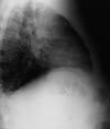
Chest radiographs are ordered.
Based on the clinical, laboratory, and radiographic findings, what is the most likely diagnosis?A. Pneumocystis pneumonia
B. Sarcoidosis
C. Pulmonary tuberculosis
D. Hemosiderosis
E. Pulmonary coccidioidomycosis (diffuse coccidioidal pneumonia)
(Answer and discussion on the next page.)
EPIDEMIOLOGY
Coccidioidomycosis is caused by the soil-dwelling dimorphic fungus Coccidioides immitis. Areas in the United States in which this fungus is endemic include the central valley of California, Arizona, Nevada, New Mexico, and western Texas. The fungus prefers the lower Sonoran life zone, which is characterized by hot summers, winter freezes, annual rainfall between 5 and 20 inches, and alkaline soil.
Approximately 100,000 cases of coccidioidomycosis occur in the United States each year. The disease has become a growing health concern, particularly in the San Joaquin Valley, because of increasing numbers of migrant farm workers; development of housing colonies (which disrupts the soil); and increasing numbers of susceptible and immunocompromised patients. Persons who travel through an area in which C immitis is endemic also become susceptible to coccidioidomycosis.
RISK FACTORS
Infection results from inhalation of the arthroconidia (airborne spores) of the fungus. Because of the organism's habitat, the incidence of disease is higher in persons exposed to dust and soil, such as oil-field workers and farmers. Persons of any age may be infected, and men are at higher risk than women.
High-risk patients include immunocompromised persons (such as those with HIV infection); those receiving immunosuppressive agents, corticosteroids, or chemotherapy; persons who have diabetes; and women in the second or third trimester of pregnancy. Certain racial groups are at higher risk for dissemination and severe disease; African Americans, Native Americans, Hispanics, and Asians (particularly those of Filipino descent) are at greater risk than whites for extrapulmonary disease.
CLINICAL MANIFESTATIONS
Most patients with coccidioidal infection experience a primary infection in the lungs; approximately 60% are asymptomatic.
Pulmonary disease. Symptoms of primary pulmonary infection include high fever with chills and rigors; profuse night sweats; generalized body aches; and severe fatigue in association with dry cough, dyspnea, or pleuritic chest pain. Arthralgia with myalgia may be seen in conjunction with rashes such as erythema nodosum or erythema multiforme. Physical examination may reveal evidence of pneumonia or pleural effusion.
Chest radiographic abnormalities are seen in up to half of all symptomatic patients. Typically, infiltrates are associated with ipsilateral hilar adenopathy. Peripneumonic pleural effusion may occur.
Most symptoms in patients with acute infection clear without treatment within 2 to 3 weeks, although it takes some patients months to fully recover. In about 5% of patients, residual pulmonary lesions or complications such as a pulmonary nodule, pulmonary cavity, or a diffuse pneumonic pattern develop. Patients with diabetes are especially at risk.
Diffuse pneumonia may extensively involve the lungs in both primary and late disease. In primary disease, diffuse pulmonary involvement may indicate multiple sites of infection that have resulted from inhalation of large numbers of arthroconidia, rather than from immunosuppression.
Diffuse pneumonia may also result from hematogenous spread. This is a rapidly progressive condition seen most commonly in immunocompromised patients; it may cause respiratory failure. Chest radiographs typically show a diffuse reticulonodular pattern throughout both lung fields.
Extrapulmonary disease. About 1% of patients are affected by extrapulmonary, or disseminated, disease attributable to hematogenous spread of the fungus. Symptoms, which usually develop several months after the initial infection, vary widely depending on the site or sites of dissemination.
Disseminated coccidioidomycosis may be nonmeningeal or meningeal. Nonmeningeal signs include cutaneous manifestations that range from papules and verrucous plaques to ulcerations. Bony lesions can affect the large bones (tibia or femur) or joints (knee, ankle, or smaller joints of the hands and feet) and cause osteomyelitis, synovitis, or joint destruction. Another common site of osteomyelitis is the vertebral column; sequelae may include diskitis, destruction of vertebral bodies, and epidural and spinal abscesses that result in various neurologic manifestations, including paraplegia with bladder and bowel involvement.
Meningeal disease, often the most lethal form of infection, causes chronic granulomatous meningitis of the basal meninges. Cerebral and cerebellar abscesses have also been reported. Usual symptoms are headaches, vomiting, vision disturbances, altered mental status, seizures with nuchal rigidity, diplopia, and cranial neuropathy. The CSF usually reveals mononuclear cell pleocytosis with elevated protein and low glucose levels; about 70% of patients may have eosinophilia. CT and MRI may show dilated ventricles with hydrocephalus and enhancement of the basal meninges.
Disseminated cocci can affect any organ. Reported sites of infection include the eye, larynx, thyroid, pericardium, peritoneum, adrenal glands, prostate, kidney, testis, and uterus. Prosthetic grafts and peritoneal shunts may also be infected.
DIAGNOSIS
A high index of suspicion is essential for prompt diagnosis. An accurate travel history is important for patients who live in areas where the fungus is not endemic.
The definitive diagnosis is established by:
- Isolation of the organism from a clinical specimen. The fungus grows rapidly on most culture mediums. Tissue specimens, including sputum and bronchiolar lavage fluid, show typical coccidioidal spherules.
- Coccidioidal antibody detection.
Two types of serologic tests are available for antibody detection:
Tube precipitin test. Tube precipitin-reacting antibodies are reported as positive IgM or IgG antibodies. IgM antibodies appear in serum within 10 days of a new infection. Thus, initial serologic studies may yield negative results. The test must be repeated in 2 weeks to establish the diagnosis.
CF test. CF antibodies are reported in titers and usually become detectable by the fourth week of infection. Changes in antibody titer generally reflect the course of the disease. CF antibodies are found in the CSF of patients with coccal meningitis.
Both tube precipitin and CF antibodies can be detected by immunodiffusion or enzyme-linked immunoassays.
MANAGEMENT
Treatment of pulmonary coccidioidomycosis involves a decision about whether intervention is necessary; the selection of antifungal agents; and a determination of whethersurgical procedures might be necessary for debridement, drainage, removal of destructive lesions, and treatment of complications (such as insertion of a ventriculoperitoneal shunt for hydrocephalus in meningitis). Treatment is recommended for symptomatic patients, those with evidence of infiltrates on chest radiographs, and those with elevated levels of CF titers.
A thorough history taking, physical examination, and laboratory and radiologic evaluation are necessary in all patients with newly diagnosed coccidioidal infection to determine the extent of disease, regardless of whether the patient is symptomatic or asymptomatic, and to identify factors that may increase the risk of complications.
Azole derivatives and amphotericin are the recommended therapeutic agents. Azole derivatives include fluconazole, itraconazole, and a newer agent, voricazole. These drugs are available in oral form and are much less toxic than amphotericin B. They are widely used in moderate to severe coccidioidomycosis. Fluconazole, the most commonly used azole, is dosed at 400 to 1000 mg/d, depending on the severity of infection. Higher doses are used in meningeal disease.
Amphotericin B (or preferably liposomal amphotericin B) is used parenterally in severe, progressive, complicated coccidioidal infections; in pregnancy (because azoles are contraindicated); in immunocompromised patients; and in those who cannot tolerate azoles or in whom azole therapy has failed. The usual dosage of amphotericin B is 0.7 to 1.0 mg/kg/d IV; the usual dosage of liposomal amphotericin B is 5 mg/kg/d IV. Treatment duration averages 6 weeks to 6 months in patients with pulmonary disease, depending on symptom resolution, radiologic clearance, and CSF titers. Patients are subsequently followed every 4 to 6 weeks until the infection has cleared; patients with disseminated disease are followed indefinitely.
In extrapulmonary meningeal disease, initial therapy involves both parenteral and intrathecal amphotericin B (because amphotericin B does not cross the blood-brain barrier), followed by a maintenance dose of azole therapy for life.
Referral to a pulmonary or infectious disease specialist is recommended for patients with complicated or disseminated disease, those who do not respond to azole therapy, and pregnant women.
References:
FOR MORE INFORMATION:
- Chiller TM, Galgiani JN, Stevens DA. Coccidioidomycosis. Infect Dis Clin North Am. 2003;17:41-57, viii.
- Einstein HE, Johnson RH. Coccidioidomycosis: new aspects of epidemiology and therapy. Clin Infect Dis. 1993;16:349-354.
- Galgiani JN. Coccidioidomycosis. West J Med. 1993;159:153-171.
- Galgiani JN, Ampel NM, Catanzaro A, et al. Practice guideline for the treatment of coccidioidomycosis. Infectious Diseases Society of America. Clin Infect Dis. 2000;30:658-661.
- Stevens DA. Coccidioidomycosis. N Engl J Med. 1995;332:1077-1082.
