- Clinical Technology
- Adult Immunization
- Hepatology
- Pediatric Immunization
- Screening
- Psychiatry
- Allergy
- Women's Health
- Cardiology
- Pediatrics
- Dermatology
- Endocrinology
- Pain Management
- Gastroenterology
- Infectious Disease
- Obesity Medicine
- Rheumatology
- Nephrology
- Neurology
- Pulmonology
A Man With Transient Dyspnea After Taking Tadalafil
A 26-year-old man presented with sudden onset of palpitations and shortness of breath after incidentally taking tadalafil. He had no other symptoms and no history of illnesses during childhood. He drank socially but denied smoking and use of illicit drugs.
Case Presentation
A 26-year-old man presented with sudden onset of palpitations and shortness of breath after incidentally taking tadalafil. He had no other symptoms and no history of illnesses during childhood. He drank socially but denied smoking and use of illicit drugs. On further questioning, he reported that 2 years ago while on a trip to a high-altitude area, he experienced episodes of excessive dyspnea. His family history was unremarkable.
Physical examination
The patient's vital signs were normal. Examination of the chest revealed diminished breath sounds on the left side. Findings from the remainder of the physical examination were unremarkable. Laboratory values were all within the normal range.
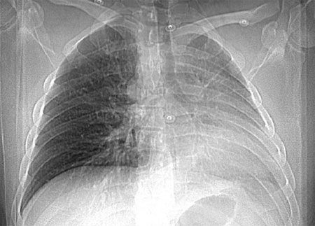
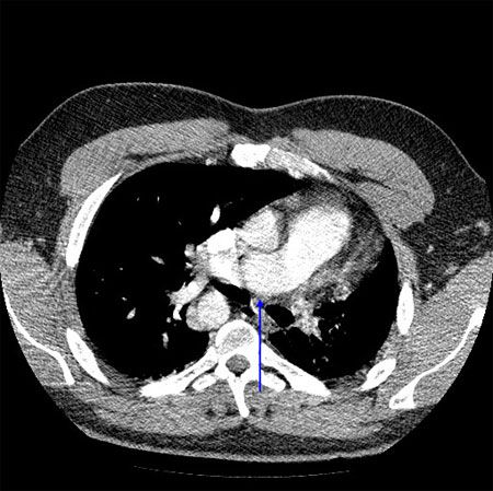
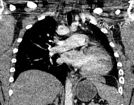
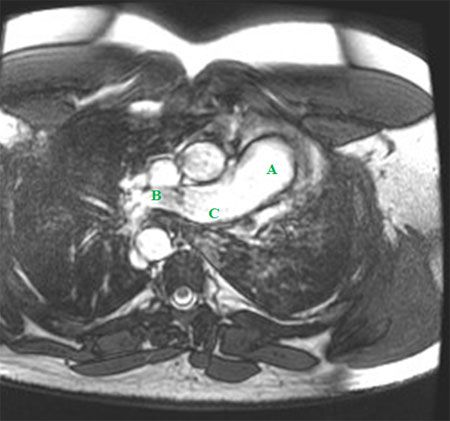
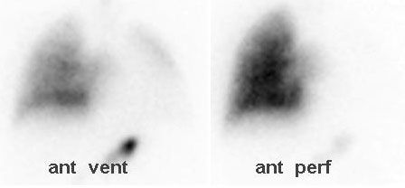
An ECG showed no significant abnormalities. A prominent right pulmonary artery, a right-sided aortic arch, cardiomegaly, and diminished left lung volumes were seen on a chest radiograph (Figure 1). An echocardiogram demonstrated normal chambers with no systolic dysfunction, no evidence of valvular or congenital heart disease, and normal pulmonary artery pressures. CT angiograms (CTAs) of the chest (Figures 2 and 3), a cardiac MRI scan (Figure 4), and a ventilation-perfusion (V/Q) lung scan (Figure 5) were also obtained. Pulmonary function tests (PFTs) revealed a moderate restrictive pattern (total lung capacity, 68% of predicted) with a mildly decreased carbon monoxide-diffusing capacity (76% of predicted) as per the ATS/ ERS task force (2005) standardization guidelines.
The differential diagnoses under consideration were left lung atelectasis, hypoplastic left lung, and congenital heart disease.
WHAT'S YOUR DIAGNOSIS?
ANSWER: Isolated unilateral absence of pulmonary artery (UAPA)
The CTAs showed complete absence of the left pulmonary artery, an enlarged right pulmonary artery, and a hypoplastic left lung (Figures 2 and 3). Isolated complete absence of the left pulmonary artery, an enlarged right pulmonary artery, a hypoplastic left lung, and a right-sided aorta were seen on the cardiac MRI scan (Figure 4). The V/Q lung scan showed hypoventilation of the left lung, with complete absence of left lung perfusion (Figure 5). These findings confirmed the diagnosis of isolated unilateral absence of pulmonary artery (UAPA).
Tadalafil is an inhibitor of phosphodiesterase type 5 (PDE-5) in smooth muscle of pulmonary and systemic vasculature via degradation of cyclic guanosine monophosphate (cGMP). Increased cGMP concentration results in vasodilation in the pulmonary bed and the systemic circulation. The latter mechanism was likely related to the patient's symptoms in the present case. The decrease in preload from systemic vasodilatation likely resulted in increased sympathetic drive and tachycardia to compensate for the decreased cardiac output. This was responsible for the transient sensation of dyspnea and palpitations. His symptoms resolved after the cessation of the drug.
The patient was subsequently discharged home. He was cautioned to avoid high-altitude areas and was warned of the dangers associated with UAPA, including hemoptysis.
Discussion
UAPA, first described in 1868 by Frantzel,1 is a rare anomaly, with an estimated prevalence of 1 in 200,000, as suggested by Bouros and colleagues. 2 Since 1978, only 119 cases have been reported.3 UAPA is often associated with congenital cardiovascular defects, such as tetralogy of Fallot, septal defect, transposition of great vessels, right-sided aortic arch, and patent ductus arteriosus.2 Cases of UAPA without any congenital defects are rarely seen. UAPA represents an embryological developmental abnormality in the left fourth and proximal sixth aortic arches. Right-sided UAPA is twice as common as left-sided UAPA,4 although the latter was noted in our patient.
Many patients with isolated UAPA who do not have cardiac anomalies have an asymptomatic clinical course and generally survive into adulthood. The workup is often prompted by abnormal findings on a chest radiograph, as demonstrated in the patient presented in this case. Earlier reports suggest that as many as 30% of UAPA patients may be asymptomatic2; however, a more recent report from 2002 of 108 patients with isolated UAPA indicated a lower percentage of asymptomatic patients (13%).4
When patients are symptomatic, they present with chest pain, pleural effusions, dyspnea, or exercise limitations (40%); recurrent pulmonary infections (37%); and/or hemoptysis (20%).4 Pulmonary hypertension has been noted in 20% to 25% of patients with UAPA and has been unmasked by predisposing factors, such as high-altitude pulmonary edema (HAPE), which our patient may have experienced during his high-altitude trip, and pregnancy. Pulmonary hypertension is the most important factor for determining survival for patients with UAPA.
In the absence of associated cardiac disease or pulmonary hypertension, exercise limitation can be defined on PFTs by a mild restrictive pattern with increased dead space ventilation to tidal volume ratio during exercise.5 The resultant hypercapnia causes bronchoconstriction in the hypoplastic lung, impaired mucociliary clearance, and diminished delivery of inflammatory cells, which likely predisposes patients to recurrent infections.2 Hemoptysis in patients with UAPA is secondary to either hypertrophied collateral vessels or the presence of a peripheral arteriovenous fistula. These anomalous collateral vessels can come from the bronchial, intercostal, subclavian, or subdiaphragmatic arteries.2,4
The chest radiograph may show ipsilateral cardiac and mediastinal displacement, ipsilateral smaller hemithorax, absent pulmonary artery shadow, ipsilateral hemidiaphragm elevation, ipsilateral absent or diminished pulmonary vascular markings, contralateral lung hyperinflation and herniation beyond the midline, or right-sided aortic arch.6
Echocardiography is useful for excluding other cardiac anomalies and for screening for pulmonary hypertension. Contrast-enhanced high-resolution CT and MRI have proved valuable as well. The advantage of combined 3-dimensional cine gradient echo MRI and magnetic resonance angiography using gadolinium is that there is no radiation. Also, findings from these studies have excellent correlation with findings from cardiac catheterization and echocardiography.6 Right-sided and left-sided heart catheterization are generally used for evaluation of pulmonary hypertension.
V/Q lung scans characteristically show complete absence of perfusion to the affected lung coupled with diminished ventilation on the wash-in and equilibrium phases but no delay in the wash-out phase of the ventilation scan.2 V/Q lung scans are typically used preoperatively to predict postoperative lung function.6 The gold standard, pulmonary angiography, should be reserved for patients who are undergoing embolization for hemoptysis or being considered for revascularization surgery.6
On fiberoptic bronchoscopy, the most frequent findings coincide with a history of recurrent pulmonary infections. It may also reveal plexuses of dilated blood vessels, mesh-like vascularization, and erythema in the bronchi on the affected side. Patients with these findings are prone to hemoptysis.2 In a few reported cases, bronchoscopy revealed mixed cylindric and saccular bronchiectasis with normal anatomy of the segmental bronchi and the contralateral lung.2 However, bronchoscopy is not routinely performed; it is used in patients who have hemoptysis or recurrent infections.
When UAPA with cardiac anomalies is diagnosed within the first year of life, it is often treated surgically. Pneumonectomy or lobectomy was performed in only 8% of the previously mentioned 119 cases of isolated UAPA, and only because the patients had recurrent hemoptysis or intractable pulmonary infections. Revascularization of hidden pulmonary arteries was performed in 7% of the patients. Massive pulmonary hemorrhage, pulmonary hypertension and right-sided heart failure, respiratory failure, and HAPE resulted in an overall mortality rate of 7%.4
When pulmonary hypertension is present in a patient with UAPA, his or her condition may be improved by revascularization of the side with the absent artery. In most cases, there is an identifiable artery at the hilum that can be demonstrated by pulmonary venous wedge angiography and may be used for revascularization. If sufficiently large hilar arteries are found, revascularization may significantly improve outcome.4 Long-term outcome for pediatric patients treated in this manner is excellent, with resolution of heart failure and pulmonary hypertension.3 If revascularization is not feasible or if pulmonary hypertension does not improve, therapeutic measures similar to those used for primary pulmonary hypertension may be helpful.
Teaching Points
1. Unilateral absence of pulmonary artery (UAPA) represents a developmental abnormality in the left fourth and proximal sixth aortic arches, often associated with various cardiovascular anomalies.
2. Patients with isolated UAPA may remain asymptomatic or present late in adulthood with exertional dyspnea, chest pain, hemoptysis, or recurrent pulmonary infections.
3. The diagnosis is confirmed with CT, MRI, V/Q lung scanning, echocardiography, or angiography.
4. Patients should be screened for pulmonary hypertension, an important predictor of survival; advised against pregnancy; and cautioned about the possibility of high-altitude pulmonary edema, hemoptysis, and recurrent pulmonary infections.
5. Therapeutic options for symptomatic patients include pneumonectomy, revascularization, and medical management of pulmonary hypertension
References:
References:
1. Frantzel O. Angeborener Defect der Rechten Lungenarterie. Virchows Arch Pathol Anat. 1868;43:420.
2. Bouros D, Pare P, Panagou P, et al. The varied manifestation of pulmonary artery agenesis in adulthood. Chest. 1995;108:670-676.
3. Atik E, Tanamati C, Kajita L, Barbero-Marcial M. Isolated unilateral pulmonary artery agenesis: evaluation of long term evolution after corrective surgery. Arq Bras Cardiol. 2006;87:423-428.
4. Ten Harkel AD, Blom NA, Ottenkamp J. Isolated unilateral absence of a pulmonary artery: a case report and review of the literature. Chest. 2002;122:1471-1477.
5. Brassard JM, Johnson JE. Unilateral absence of a pulmonary artery. Data from cardiopulmonary exercise testing. Chest. 1993;103:293-295.
6. Griffin N, Mansfield L, Redmond KC, Dusmet M, et al. Imaging features of isolated unilateral pulmonary artery agenesis presenting in adulthood: a review of four cases. Clin Radiol. 2007;62:238-244.
