- Clinical Technology
- Adult Immunization
- Hepatology
- Pediatric Immunization
- Screening
- Psychiatry
- Allergy
- Women's Health
- Cardiology
- Pediatrics
- Dermatology
- Endocrinology
- Pain Management
- Gastroenterology
- Infectious Disease
- Obesity Medicine
- Rheumatology
- Nephrology
- Neurology
- Pulmonology
Livedo Reticularis
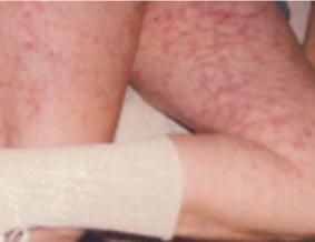
A 62-year-old woman was seen prior to cholecystectomy. She had no cardiac or pulmonary disorders. Examination was unremarkable other than a purplish-reddish, lace-like pattern on the thighs and forearms.

A 62-year-old woman was evaluated in preparation for cholecystectomy. She had no cardiac or pulmonary disorders. The physical examination was unremarkable other than a purplish red lace-like pattern on the skin. The mottling was most prominent on the thighs (Figure) and forearms. She had been experiencing the rash for 3 years and said that it becomes more pronounced when she is exposed to cold. The lesions caused no itching or pain.
This is a classic presentation of livedo reticularis, a chronic vascular disorder in which constriction of cutaneous blood vessels leads to the characteristic mottled or net-like discoloring on large areas of the legs or arms and, less frequently, the trunk. The condition is aggravated by cold exposure. Livedo reticularis can be primary or secondary. The primary, or idiopathic, form occurs in adults, most often women, in their 30s and 40s. Typically this form causes no symptoms and needs no treatment. Patients usually seek medical attention for cosmetic reasons.
Secondary livedo reticularis may provide a dermatologic clue to a more serious systemic condition (see below) that is often characterized by vessel wall disease or intravascular obstruction. Regardless of the primary pathology, livedo reticularis signals a common abnormality-impaired blood flow in the cutaneous arteriolar cones. Deoxygenated blood stagnates and causes cyanotic discoloration at the anastomoses between cones. The resultant mottling reflects the vascular anatomy of normal skin.
In the appropriate setting, a complete blood cell count and anti-nuclear antibody testing should be performed with the objective of diagnosing and treating the underlying condition. In most cases, however, this distinctive rash has few if any sequelae. In the absence of any other signs and symptoms of disease, past or present, patients should be reassured and advised to avoid cold environments.
Causes of livedo reticularis
1. Idiopathic
2. Lymphoproliferative malignancies-may provoke livedo reticularis via hyperviscosity, hyperproteinemia, or thrombocytosis.
3. Collagen vascular disorders-systemic lupus erythematosus, rheumatoid arthritis, polyarteritis nodosa, dermatomyositis, cryoglobulinemia, and primary antiphospholipid antibody syndrome.
4. Hereditary protein C deficiency and antithrombin III deficiency
5. Embolic phenomenon-ie, cholesterol emboli syndrome, an often catastrophic event that occurs after invasive radiologic procedures like cardiac catherterization.
6. History of ischemic cerebrovascular events-ie, Sneddon’s syndrome
The lace-like pattern of livdeo reticularis is similar to that seen in erythema ab igne (Figure) The latter is associated with heat exposure.
References:
Sources:
Anderson RA, D'Cruz D, Merry P, et al. Cryoglobulins, anticardiolipin antibodies, and livedo reticularis. J Rheumatol. 1992;19:826.
Sneddon I. Cerebrovascular lesions and livedo reticularis. Br J Dermatol. 1965;77:180-185.
Gibbs M, English J, Zirwas M. Livedo reticularis: an update. J Am Acad Dermatol. 2005;52:1009-1019.
Oral PCSK9 Inhibitor Enlicitide Meets All End Points in Phase 3 Hypercholesterolemia Trial
September 2nd 2025At week 24, patients receiving once-daily enlicitide demonstrated statistically significant and clinically meaningful reductions in low-density lipoprotein cholesterol compared with placebo.
