- Clinical Technology
- Adult Immunization
- Hepatology
- Pediatric Immunization
- Screening
- Psychiatry
- Allergy
- Women's Health
- Cardiology
- Pediatrics
- Dermatology
- Endocrinology
- Pain Management
- Gastroenterology
- Infectious Disease
- Obesity Medicine
- Rheumatology
- Nephrology
- Neurology
- Pulmonology
Lethargy, Confusion, and Constipation in an Older Woman
A 77-year-old woman is brought for evaluation by her family. The patient had previously been alert and active; however, for the past week, she has been difficult to arouse and, when awake, has been delusional and has behaved abnormally. In addition, for the past 2 weeks, she has complained of abdominal discomfort related to constipation.
A 77-year-old woman is brought for evaluation by her family. The patient had previously been alert and active; however, for the past week, she has been difficult to arouse and, when awake, has been delusional and has behaved abnormally. In addition, for the past 2 weeks, she has complained of abdominal discomfort related to constipation.
HISTORY
The patient has no history of heart disease, diabetes, or hypertension. She had a 40-pack-year smoking history but quit 2 years earlier when mild chronic obstructive pulmonary disease was diagnosed. She has been coughing more than usual in recent months but without producing sputum or blood. Her medications include montelukast sodium and a statin.
PHYSICAL EXAMINATION
The patient is disoriented. Heart rate is 108 beats per minute; respiration rate, 20 breaths per minute; blood pressure, 105/60 mm Hg; and oxygen saturation measured by pulse oximetry, 93% on room air. Mucous membranes are quite dry. A 3-cm hard lymph node is palpable in the left supraclavicular fossa. Examination of the chest reveals decreased breath sounds and a few wheezes but no consolidation; the heart is normal. Neurological examination reveals disorientation but no focal neurological findings.
LABORATORY AND IMAGING RESULTS
Hemoglobin level is 10.5 g/dL; white blood cell count, 8100/μL; and platelet count, 517,000/μL. Creatinine level is 2.3 mg/dL, and blood urea nitrogen level is 26 mg/dL. Albumin level is 2.9 g/dL, with a total protein level of 5.9 g/dL. Serum glucose level is 110 mg/dL, and serum calcium level is 14.8 mg/dL. A chest radiograph reveals fullness of the left hilum of the lung.

(answer on next page)
CORRECT ANSWER: C
Considered individually, many of the clinical manifestations of hypercalcemia are nonspecific. However, when confusion, lethargy, fatigue, nausea, vomiting, constipation, and dehydration are clustered together, hypercalcemia is the likely cause. Suspected hypercalcemia can be confirmed by measuring the serum calcium level (or, for greater accuracy, the ionized calcium level).
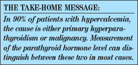
Causes of hypercalcemia. Hypercalcemia can have a variety of causes; however, malignancy and primary hyperparathyroidism account for at least 90% of cases. Thus, evaluation of a patient with hypercalcemia must include prompt investigation of these two possibilities.1
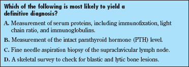
Role of PTH measurement. A normal or high intact PTH level suggests primary hyperparathyroidism. A suppressed level suggests malignancy. This patient’s intact PTH level was very low (19 pg/mL), which excluded primary hyperparathyroidism. Such a result was anticipated, given the presence of 2 clues that strongly suggested malignancy (the patient’s abnormal chest radiograph and the grossly abnormal supraclavicular lymph node). However, the finding of a low PTH level still does not point to a specific diagnosis; a neoplasm workup is required, and choice B is thus incorrect.
Diagnosing malignant causes. Neoplasms commonly associated with hypercalcemia include squamous cell cancers of the upper respiratory-digestive tract, breast cancer, myeloma, and renal cancers.2 These neoplasms are typically large, advanced, and quite evident. Other endocrine syndromes (eg, multiple endocrine neoplasia) can also be associated with hypercalcemia, and their clinical manifestations can be more subtle.

Although an exhaustive battery of radiological studies is routinely ordered when malignancy is suspected, this woman’s abnormal chest radiograph and firm, enlarged supraclavicular node obviate the immediate need for many such studies. Enlarged supraclavicular nodes are essentially always abnormal rather than reactive.
The criteria for lymph node biopsy include: presence of enlargement for more than 1 month, size greater than 2 cm, or worrisome location (eg, in the supraclavicular node).2 The selection of a biopsy technique depends on the patient’s age. Lymphoma is the most common malignancy in patients younger than 40 years; thus, a full extraction is preferred in these patients to provide sufficient tissue to determine whether lymphoma is present and, if it is, what type. In patients older than 40 years, adenocarcinoma is statistically more likely, and fine needle aspiration is an appropriate diagnostic technique. In this patient, a fine needle aspiration biopsy of the supraclavicular lymph node (choice C) will very probably reveal lung cancer, given her history of heavy smoking, new cough, and chest film abnormality.
Another common cause of neoplastic hypercalcemia is plasma cell disease, in which plasma cell production of osteoclast-activating factor results in elevated blood calcium levels.3 The workup for plasma cell disease involves measurement of the serum proteins mentioned in choice A. Patients with plasma cell disease frequently have significant skeletal involvement, with classic lytic lesions. No such lesions were evident on this patient’s chest radiographs; moreover, her total protein level was not abnormally elevated. Thus, the preponderance of the clinical findings here point to lung rather than plasma cell disease, and choice A is not correct.
A skeletal survey (choice D) is always a good idea in a patient with hypercalcemia. Such a study may reveal typical radiological patterns-the purely lytic lesions of myeloma (just mentioned) or the blastic metastases of prostate or breast cancer. However, such studies do not yield a tissue diagnosis. When tissue for diagnosis is easily obtainable, as in this patient, proceeding directly to biopsy will result in a diagnosis more quickly.
Outcome of this case. The patient’s hypercalcemia was treated initially with aggressive intravenous saline hydration and pamidronate. By the next morning, her calcium level was 12.6 mg/dL and her mental status had normalized. On the third hospital day, a fine needle aspiration biopsy of the supraclavicular lymph node revealed squamous cell carcinoma. Bronchoscopy confirmed a bronchogenic primary tumor in the left lobar bronchus. Therapy with pamidronate was continued while treatment options targeting the carcinoma were evaluated. Her prognosis is extremely guarded because hypercalcemia in patients with cancer is an ominous sign, associated with a mortality of about 50% within 30 days.4
References:
REFERENCES:1. Carroll MF, Schade DS. A practical approach to hypercalcemia. Am Fam Physician. 2003;67:1959-1966.
2. Stewart AF. Clinical practice. Hypercalcemia associated with cancer. N Engl J Med. 2005;352:373-379.
3. Kyle RA, Rajkumar SV. Multiple myeloma. N Engl J Med. 2004;351:1860-1873.
4. Ralston SH, Gallagher SJ, Patel U, et al. Cancer-associated hypercalcemia: morbidity and mortality: clinical experience in 126 treated patients. Ann Intern Med. 1990;112:499-504.
Obesity Linked to Faster Alzheimer Disease Progression in Longitudinal Blood Biomarker Analysis
December 2nd 2025Biomarker trajectories over 5 years in study participants with AD show steeper rises in pTau217, NfL, and amyloid burden among those with obesity, highlighting risk factor relevance.
