- Clinical Technology
- Adult Immunization
- Hepatology
- Pediatric Immunization
- Screening
- Psychiatry
- Allergy
- Women's Health
- Cardiology
- Pediatrics
- Dermatology
- Endocrinology
- Pain Management
- Gastroenterology
- Infectious Disease
- Obesity Medicine
- Rheumatology
- Nephrology
- Neurology
- Pulmonology
Late Infection of Ruptured Silicone Breast Implant for Breast Augmentation
A 66-year-old woman presented with a 2-day history of acute onset of redness, pain, and swelling of the right breast.

A 66-year-old woman presented with a 2-day history of acute onset of redness, pain, and swelling of the right breast. She was nauseated and had a temperature of 103.9°F (39.5°C). She had no history of trauma or recent illness. Her medical history included silicone breast augmentation in the 1970s, cervical disc surgery, mild asthma, and a history of pulmonary atypical mycobacterial infection 5 years before this visit. She had received regular screening mammograms, the most recent of which was 10 months earlier. Extracapsular silicone in the left breast had first been noted in 2007. The patient reported no other previous problems with her breast implants and was satisfied with her appearance.

Aside from fever, other vital signs were normal. The patient had bilateral palpable sub-glandular silicone implants with Baker Grade 3 capsular contracture (ie, breast firm to the touch with visible breast deformity) of the right breast. There were no masses and no axillary lymphadenopathy. The right breast was swollen, erythematous, and tender, with an intact inframammary scar. Results of ultrasonography showed no fluid collection or abscess.
A course of oral amoxicillin clavulanate was initiated for presumed cellulitis. After 48 hours of oral antibiotic treatment, the fever resolved but the erythema did not improve. The patient was admitted to the hospital and treated with intravenous vancomycin for 4 days. Nevertheless, the breast remained erythematous and tender, and the patient was advised to have the implant removed.
An incision was made in the existing inframammary scar. The implant had ruptured and the implant cavity was filled with purulent material. The pus was drained and capsule excised. All implant material was removed. The site was irrigated and sutured over a closed-suction drain. The implant in the left breast was also removed during the same operation, at the patient’s request.
Vancomycin was continued for 48 hours after surgery until blood cultures revealed Staphylococcus aureus resistant only to penicillin and tetracycline. Analyses of acid-fast bacilli cultures were ordered in light of the patient’s history of atypical mycobacterial infection, but results were negative. The patient was discharged on a 14-day course of oral levofloxacin and rifampin. She continued to heal and the infection resolved.
Breast augmentation is the most common type of cosmetic surgery elected by women in the United States. In 2010, there were a reported 318,123 augmentation procedures performed.1 Thus, primary care physicians should become familiar with the potential complications they may see.
There are 4 FDA-approved breast implant brands marketed in the US for augmentation and reconstruction. They are either filled with saline or silicone gel.2 Infections associated with breast implants are uncommon-only 1% to 2% in most reports.3,4 The majority of these occur in the immediate postoperative period. Infection rarely develops in women years after breast augmentation. If it does, treatment is similar to that for an acute infection caused by any type of foreign body. The implant is removed to allow the infection to resolve.
Breast tissue has endogenous bacterial flora derived from the skin. The organisms include coagulase negative staphylococci, diphtheroids, lactobacilli, propionibacterium, and alpha hemolytic streptococci.5 The surface of an implant may serve as a scaffold for growth of this endogenous flora into a biofilm. A biofilm is an aggregate of microbes that attaches to a solid surface and is encased by an exopolysaccharide matrix. The environment is self-sustaining.6 Tissue cultures taken at a mean of 9 years after breast implantation show subclinical infection in a significant number of patients with clinically evident capsular contracture,7 as seen in this patient.
A survey of patients with early and late mammary implant infections found that diabetes, smoking, and obesity do not significantly increase the risk for infection, but that a wide range of conditions do. These include skin atrophy and scarring, pregnancy, preceding lactation, and vigorous exercising, massage, and postsurgical trauma.8 The rate of infection is also lower in women who have cosmetic augmentation mammoplasty than among those who have breast implantation after mastectomy for cancer.9
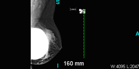
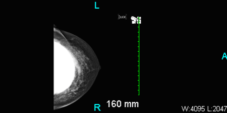
Breast augmentation with implants also can make mammography challenging.10 Additional ultrasonographic images are usually needed to detect pathology. But, as seen in this case, implant material may effectively obscure a lesion.
Failure of an infection to respond to standard antibiotic treatment may force removal of breast implants. In fact, infections associated with different types of surgical implants are character- istically antibiotic-resistant.6 Although it is rare to see an implant infection many years-or, in this case, decades-after the original procedure, cases are documented.11 This case highlights a very uncommon complication of a very common cosmetic procedure. It reminds us that infection secondary to breast augmentation should not be ruled out, even when the timeframe suggests it is highly unlikely. The case was managed in collaboration with plastic surgery, breast clinic, and infectious disease.
Figure: Ultrasound of right breast showing subglandular implants. No definite abscess seen.
References:
1. American Society of Aesthetic Plastic Surgery. Quick Facts: highlights of the ASAPS 2010 statistics on cosmetic surgery. (Accessed June 20, 2011, at:
.)
2. US Food and Drug Administration. Medical Devices; Labeling for Breast Implants. http://www.fda.gov/MedicalDevices/ProductsandMedicalProcedures/ImplantsandProsthetics/BreastImplants/ucm063743.htm. Accessed June 22, 2011.
3. Hvilsom, G et al. Local complications after cosmetic breast augmentation. Plast Reconstr Surg. 2009;124:919-925.
4. Codner, M et al. A 15 year experience with primary breast augmentation. Plast Reconstr Surg. 2011;12:1300-1310.
5. Thornton JW et al. Studies on the endogenous flora of the human breast. Ann Plast Surg. 1988;20:39-42.
6. Mah T-F C, O’Toole GA. Mechanisms of biofilm resistance to antimicrobial agents. TRENDS in Microbiology. 2001;9:34-39.
7. Pajkos et al. Detection of subclinical infection in significant breast implant capsules. Plast Reconstr Surg. 2003;111:1605
8. Brand KG et al. Infection of mammary prostheses: a survey and the question of prevention. Ann Plast Surg. 1993;30:289-295.
9. Gabriel SE, Woods JE, O’Fallon WM, et al. Complications leading to surgery after breast implantation. N Engl J Med. 1997;336:677-682.
10. Steinbach BG, et al. Mammography: breast implants-types, complications, and adjacent breast pathology. Curr Probl Diagn Radiol. 1993;22:39-86.
11. Ablaza V, LaTrenta G. Late infection of breast prosthesis with Enterococcus avium. Plast Reconstr Surg. 1998;102:227-230.
Obesity Linked to Faster Alzheimer Disease Progression in Longitudinal Blood Biomarker Analysis
December 2nd 2025Biomarker trajectories over 5 years in study participants with AD show steeper rises in pTau217, NfL, and amyloid burden among those with obesity, highlighting risk factor relevance.
