

- Clinical Technology
- Adult Immunization
- Hepatology
- Pediatric Immunization
- Screening
- Psychiatry
- Allergy
- Women's Health
- Cardiology
- Pediatrics
- Dermatology
- Endocrinology
- Pain Management
- Gastroenterology
- Infectious Disease
- Obesity Medicine
- Rheumatology
- Nephrology
- Neurology
- Pulmonology
Large Basal Cell Carcinoma
The lesion’s location, along with visible telangiectasia on the lesion’s surface, suggest a basal cell carcinoma.


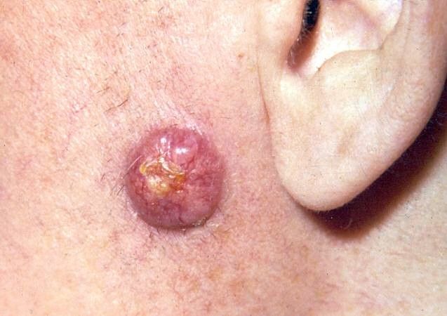
A gradually enlarging nodule on the pre-auricular facial skin developed in a 68-year-old man who was otherwise in good health. The lesion was asymptomatic. There was no regional adenopathy.
Key point: The lesion’s location, along with visible telangiectasia on the lesion’s surface, suggest a basal cell carcinoma. Biopsy verified this tentative diagnosis.
Treatment: Because of the lesion’s size (2 cm x 2 cm) and location (near branches of the facial nerve), Moh’s surgery with microscopic control was performed, rather than deep excision.
Note: While basal cell carcinoma only rarely metastasizes, it can cause important anatomical disruption in select areas and may require very careful removal.


