- Clinical Technology
- Adult Immunization
- Hepatology
- Pediatric Immunization
- Screening
- Psychiatry
- Allergy
- Women's Health
- Cardiology
- Pediatrics
- Dermatology
- Endocrinology
- Pain Management
- Gastroenterology
- Infectious Disease
- Obesity Medicine
- Rheumatology
- Nephrology
- Neurology
- Pulmonology
Iritis Caused by Neurosyphilis
Most cases of “pink eye” are caused by infectious, or occasionally allergic, conjunctivitis. Occasionally, however, a rare and more serious condition-such as a corneal ulcer or iritis-may masquerade as conjunctivitis, as in this patient.

A 43-year-old man presents with left eye irritation that has been present for about 2 weeks. He denies trauma, headache, eye discharge, fever, cough, and vomiting, but does have some mild photophobia and slightly blurred vision that does not seem to clear when he blinks. He has had similar symptoms twice in the past 2 years and was told that he had conjunctivitis both times. He took antibiotic drops, which resolved the symptoms after each episode.
Currently, a coworker is taking medication for “pink eye.”
The patient wears soft contact lenses, but denies that he kept them in while sleeping. His past medical history is unremarkable except for hypertension, for which he takes metoprolol. He denies any prior surgeries, including any eye surgeries. There are no rashes.
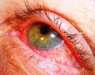
His vital signs are all normal. His head shows no swelling or temporal artery tenderness. There is no proptosis or eye pain with motion. The lids and lashes appear normal. The right pupil appears normal, but the left pupil is slightly smaller and reacts more slowly. There is also mild bilateral photophobia and consensual photophobia. The right eye is clear, but the left eye shows injection without discharge (Figure).
DISCUSSION
In this patient, conjunctivitis was not misdiagnosed a third time. The patient was referred to an ophthalmologist and was seen the next day. It was determined that the iritis was caused by neurosyphilis. He was treated with parenteral penicillin and topical corticosteroids.
Most cases of red eye or “pink eye” are caused by infectious, or occasionally allergic, conjunctivitis. Occasionally, however, a rare and more serious condition-such as a corneal ulcer or iritis-may masquerade as the more benign condition, conjunctivitis.
About Iritis
Distinguishing characteristics of iritis include consensual photophobia, limbal flush, cell and flare, and occasionally posterior synechiae.
• Consensual photophobia is pain elicited in the affected eye when light is shined into the unaffected eye. Although ipsilateral photophobia may occur in many eye conditions, consensual photophobia is rare except in iritis. It may be absent, however, in chronic iritis.
• Limbal flush is an intense injection in the bulbar conjunctiva immediately adjacent to the cornea, which occurs in conditions that affect the iris or cornea. It is can be seen clearly in the image for this patient. It should not occur in conditions limited to the conjunctiva (ie, in conjunctivitis there is typically limbal sparing, or a band of decreased or absent erythema).
• Cell and flare are the manifestations of inflammation seen in the anterior chamber on high magnification with the slit lamp and were noted in the examination of this patient. This is best seen when the light beam is narrowed and shortened so as to minimize glare off the cornea.
• Posterior synechiae are bands of pigmented scar tissue that connect the posterior surface of the iris to the anterior surface of the lens in chronic or recurrent iritis. They may limit the excursion of the pupil by tethering, as in this case. (The glare in the Figure may obscure the synechiae.)
Finally, it is crucial to measure intra-ocular pressure and visual acuity since they may be affected in iritis.
Referral and treatment
A patient with suspected iritis should be referred to an ophthalmologist. Treatment of iritis hinges on determining its primary cause. Although most cases are idiopathic, other causes are not uncommon-especially when the iritis is recurrent, as it was in this patient.
Most non-idiopathic causes of iritis are either caused by trauma, infection, or a chronic inflammatory condition (Table).
Initial treatment usually involves a topical cycloplegic agent, such as homatropine, and a topical corticosteroid once infection has either been ruled out or is diagnosed and simultaneously treated. Phone consultation and /or next day follow-up with the ophthalmologist should be arranged when the patient is seen in the clinic or emergency department.
Complications of iritis include synechiae, secondary glaucoma, and loss of vision from involvement of the uvea, which nourishes the retina. Synechiae are usually due to chronic or recurrent iritis; these do not require emergency treatment, but usually respond to a combination of steroid, cycloplegic, and sympathomimetic eye drops. Elevated intra-ocular pressure, however, should be treated immediately with medication for glaucoma. Any significant loss of vision leads to concern for more severe disease and therefore necessitates urgent follow-up or immediate evaluation by the ophthalmologist.
