- Clinical Technology
- Adult Immunization
- Hepatology
- Pediatric Immunization
- Screening
- Psychiatry
- Allergy
- Women's Health
- Cardiology
- Pediatrics
- Dermatology
- Endocrinology
- Pain Management
- Gastroenterology
- Infectious Disease
- Obesity Medicine
- Rheumatology
- Nephrology
- Neurology
- Pulmonology
HSV Esophagitis
The most frequently encountered cause of infection of the esophagus is Candida. Among viral causes HSV is the most common, followed by CMV.
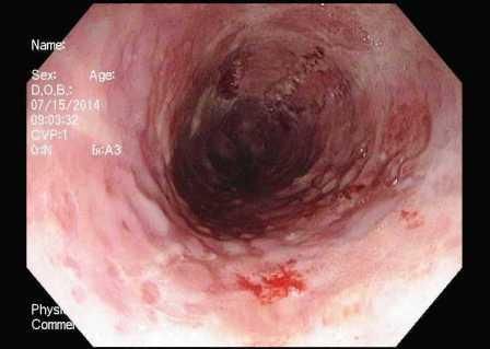
Figure. Upper endoscopy (click on figure to enlarge)

A 28-year-old man presents with a 1-week history of odynophagia. He reports being in his usual state of health before the onset of this symptom. He denies fevers, chills, or upper respiratory tract symptoms. His gastrointestinal review of systems is otherwise unremarkable. On examination, he appears uncomfortable when asked to swallow. Results from his oropharyngeal examination are normal. He undergoes upper endoscopy for further evaluation (Figure). Results of biopsies reveal Cowdry inclusion bodies and ground glass nuclei.
1. What is the most common cause of infectious esophagitis?
2. From where specifically should esophageal biopsies be taken?
3. What is the preferred treatment for this patient’s condition?
Answers and Discussion
The most frequently encountered cause of infection of the esophagus is Candida, and many healthy patients can be treated with an empiric course of fluconazole, especially if white plaques are noted on the tongue or pharynx. Among the viral causes of esophagitis, herpes simplex virus (HSV) is the most common, followed by cytomegalovirus (CMV).
Although HSV esophagitis is much more common in immunosuppressed individuals, it can occur in healthy persons. Endoscopically, the ulcers are described as shallow-appearing or as punched-out lesions, as seen in the Figure. Biopsies should be taken from both the center and periphery of the ulcer. For HSV, the viral cytopathic features are usually present adjacent to the ulcer, because the virus infects the squamous epithelial cells. On histopathology, Cowdry type A inclusion bodies, which are eosinophilic clusters, can be seen in cells infected with HSV.
In contrast, CMV infects endothelial cells, which are commonly found in the ulcer base. For these reasons, biopsies of esophageal ulcers should be taken from near the border (HSV) and in the center (CMV) of the lesion. As for treatment, the majority of cases of HSV esophagitis are self-limited. Topical lidocaine can reduce symptoms of odynophagia. In patients who are immunocompromised, a 10-day course of oral acyclovir can be prescribed. On the other hand, the treatment for CMV esophagitis is ganciclovir.
Clinical Tips for Using Antibiotics and Corticosteroids in IBD
January 5th 2013The goals of therapy for patients with inflammatory bowel disorder include inducing and maintaining a steroid-free remission, preventing and treating the complications of the disease, minimizing treatment toxicity, achieving mucosal healing, and enhancing quality of life.
