- Clinical Technology
- Adult Immunization
- Hepatology
- Pediatric Immunization
- Screening
- Psychiatry
- Allergy
- Women's Health
- Cardiology
- Pediatrics
- Dermatology
- Endocrinology
- Pain Management
- Gastroenterology
- Infectious Disease
- Obesity Medicine
- Rheumatology
- Nephrology
- Neurology
- Pulmonology
Hemorrhagic Pituitary Apoplexy in Pregnancy: A Retrospective Radiologic Diagnosis
A 38-year-old woman became pregnant during the time she was undergoing evaluation for a pituitary adenoma. Here, see the case unfold.
Figure A1. Coronal T1-weighted image showing hyperintense pituitary macroadema, 2.2 x 1.8 x 1.5 cm (arrow), consistent with hemorrhagic pituitary macroadema. Note extension into left cavernous sinus abutting on left internal carotid artery and deforming optic chiasm.
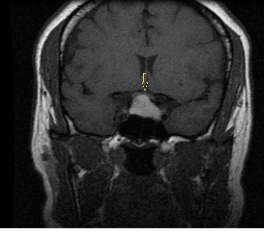
Figure A2: Axial T2-weighted image showing a fluid-fluid level consisting of a hypointense bottom layer and a hyperintense upper layer (red arrow) depicting a hemorrhagic fluid debris level.
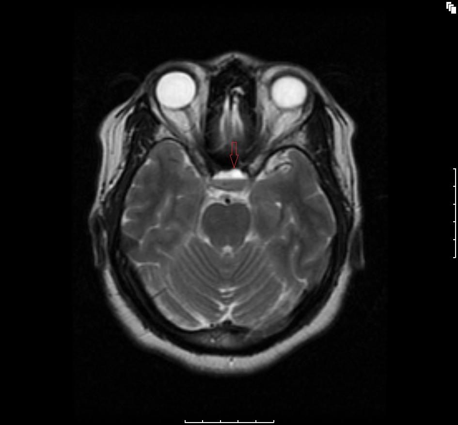
Figure A3: Axial flair T2-weighted 3 planar image showing a hypointense bottom layer and a hyperintense upper layer (green arrow) consistent with hemorrhagic pituitary macroadenoma.
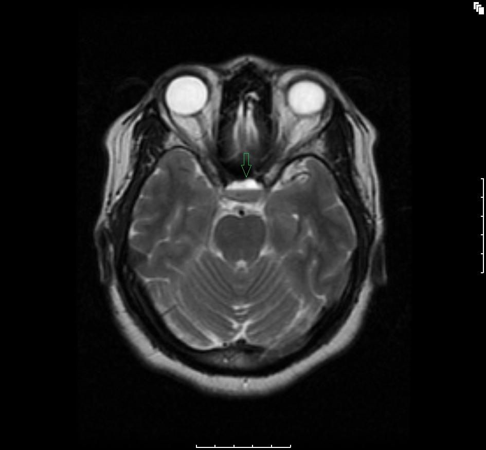
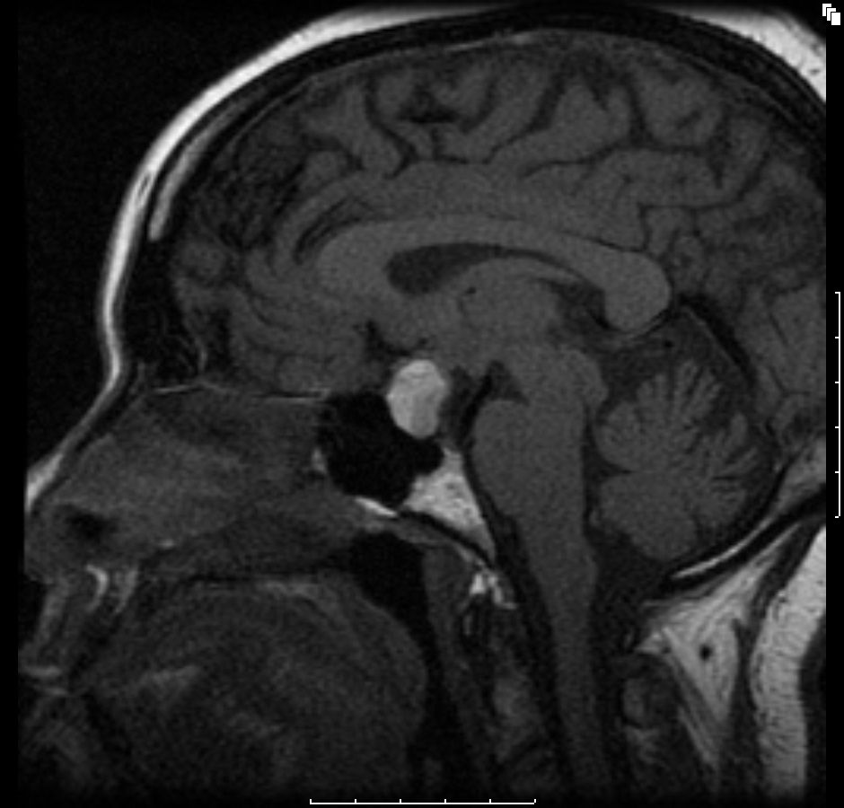
Figure A4: Sagittal T1-weighted image showing a macroadenoma in the sella extending superiorly (blue arrow), with an area of relatively low signal intensity in the posterior pituitary.
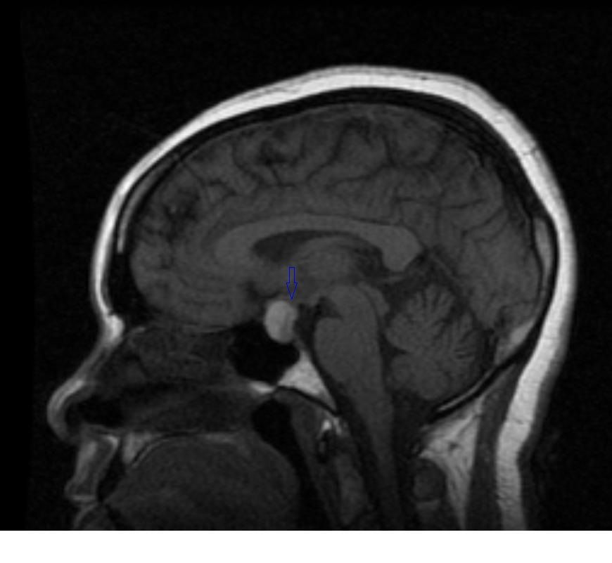
Figure B1. Coronal T1-weighted image.
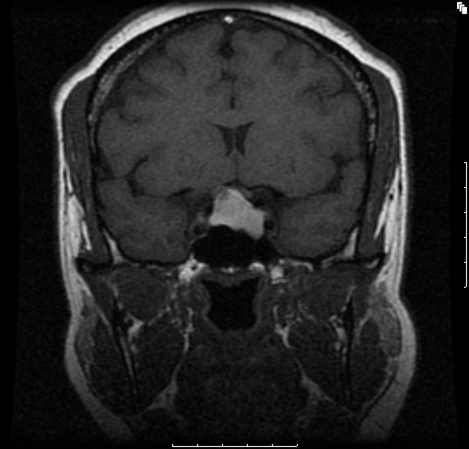
Figure B2. Axial T2-weighted image.
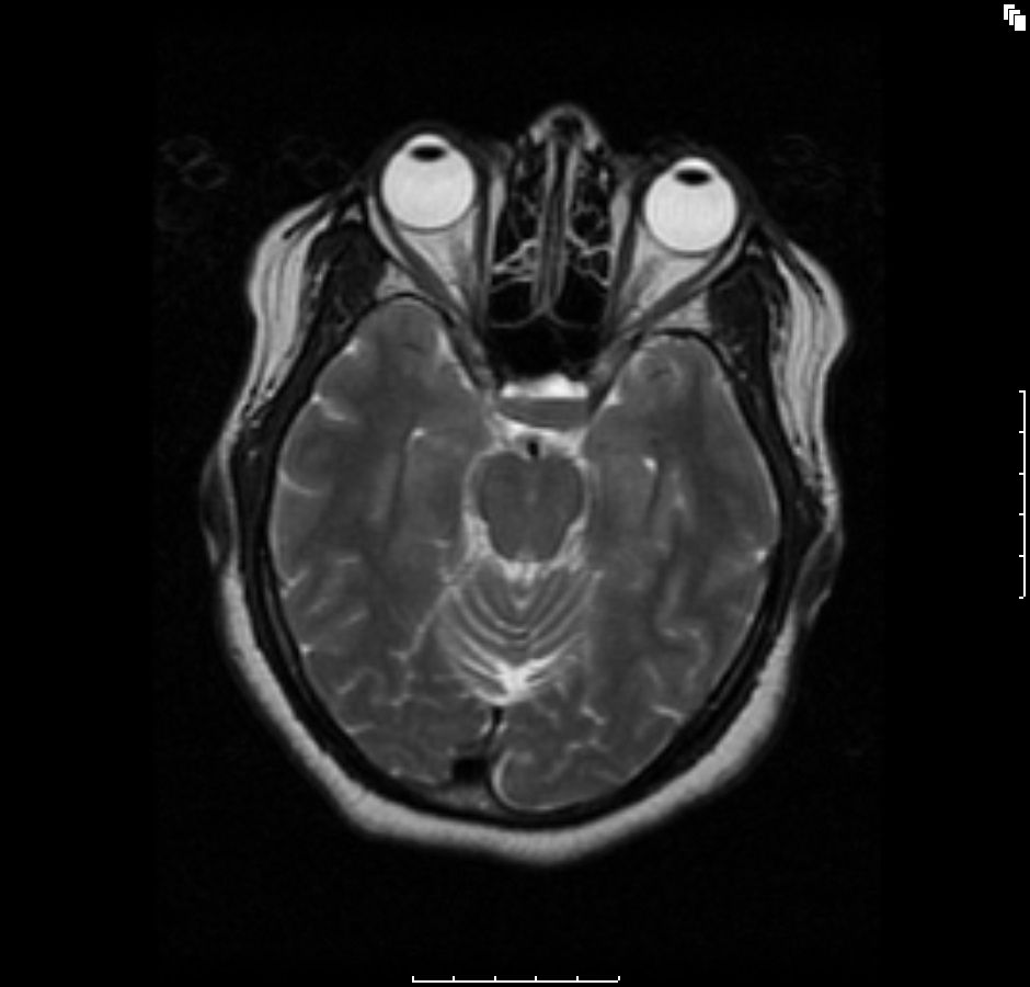
Figure B3. Axial flair T2-weighted 3 planar image.
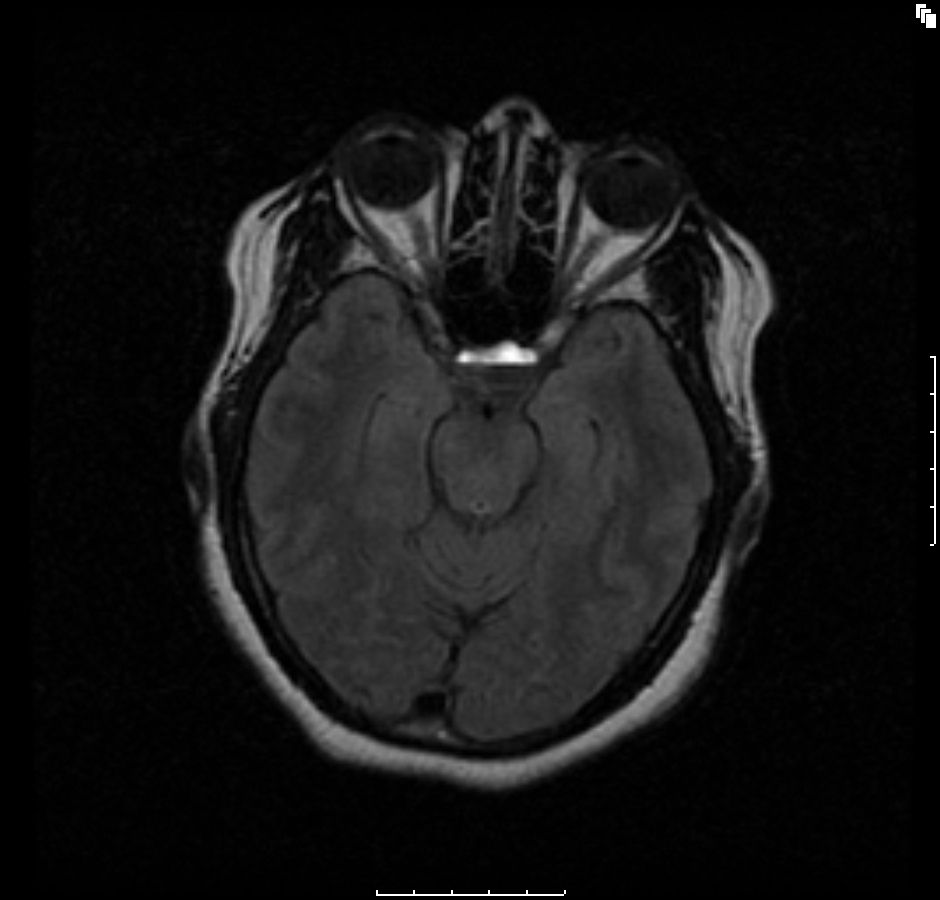
Figure B4. Sagittal T1-weighted image.
A 38-year-old woman became pregnant during the time she was undergoing evaluation for a pituitary adenoma. Three months before learning of her pregnancy, she had been seen in the endocrine clinic with complaints of galactorrhea and irregular menstrual cycles. Her laboratory workup had revealed elevated prolactin levels; a pregnancy test at the time was negative. Treatment with bromocriptine was started. There was considerable delay in obtaining initial brain MRI scans because the patient missed several appointments.
The initial brain scans were obtained 6 months after her initial diagnosis (3 months into her pregnancy) and revealed a hemorrhagic pituitary macroadenoma (Figures A1-4). The patient recollects that a week before the scan, she suffered an episode of severe headache with visual disturbance of the right eye. She did not seek medical attention at the time but did keep an appointment for the brain imaging the following week. At the time of the imaging, she reported that her symptoms had fully resolved.
She remained asymptomatic at her clinic follow-up 2 weeks after the episode, and there were no objective focal neurologic deficits. Full ophthalmologic assessment was unremarkable, with no visual field defects. She remained asymptomatic without neurologic deficits and, so far, has no features of hypopituitarism. Repeated scans performed 6 months into the patient’s pregnancy (Figures B1-4; cuts are equivalent to A series) did not show any significant interval changes. She gave birth at 9 months uneventfully and is being followed up regularly for signs and symptoms of hypopituitarism.
Discussion
Pituitary tumor apoplexy is an uncommon, acute, life-threatening neurosurgical emergency. The true incidence and prevalence is unknown but its incidence is estimated at 7.3% in patients with pituitary adenomas1 and is seen in 0.5% to 17% of patients who have had pituitary tumors surgically removed.2 The estimated male to female ratio ranges from 1:1 to 2:1.1,2 The usual age range is 37 to 57 years. The mortality rate associated with pituitary apoplexy is as high as 12.5%.2
The triggering event is sudden vascular compromise of the pituitary gland related to either hemorrhage or infarction, and there is usually an underlying pituitary adenoma. Acute neurologic impairment follows and is characterized by sudden headache, visual symptoms, altered mental status, and hormonal imbalance. These sequelae are the result of pressure from acute enlargement of an adenomatous or less commonly a non-adenomatous pituitary gland as well as the acute functional compromise of the pituitary gland. Surprisingly, a number of patients are asymptomatic or present subacutely with mild symptoms. Some predisposing factors to pituitary apoplexy include pregnancy, estrogen therapy, treatment with bromocriptine, head trauma, cardiac surgery, bilateral aderenalectomy, pituitary irradiation, anticoagulant therapy, coagulopathy, and endocrine stimulation tests.1 Our patient was pregnant and also had received bromocriptine when suspicion for pituitary tumor was high.
Management
Acute management is usually by medical stabilization, high-dose corticosteroids, other appropriate hormone supplementation, and urgent or interval surgical intervention. The management modality, however, depends largely on the clinical presentation. In patients with mild stable signs or who present subacutely after resolution of acute symptoms as our patient did, conservative management with close observation is usual. Treatment and long-term follow-up are carried out by a multidisciplinary team typically composed of neurosurgeons, endocrinologists, ophthalmologists, internists, and radiologists among other health care team members. Monitoring of pituitary hormones, visual acuity and field testing, and serial pituitary imaging by MRI every 3 to 6 months until hemorrhage resolves are recommended as initial follow-up.
Visual compromise from elevated intracranial pressure is the primary acute complication and includes optic neuritis and ophthalmoplegia with secondary ptosis and extraocular muscle paralysis.
The natural history of pituitary tumor apoplexy following conservative management includes spontaneous resolution of hemorrhage over time, pituitary shrinkage with ensuing empty-sella syndrome, and long-term sequelae of partial or complete hypopituitarism.
In one study, 87% of patients with prolactinomas had complete resolution of their hemorrhage within 26.6 ± 23.3 (mean ± SD) months.3
Patients should be well educated on the complications and sequelae and the need for continued follow-up and long-term management. We will follow our patient with serial pituitary imaging.
References:
- Möller-Goede DL, Brändle M, Landau K, et al. Pituitary apoplexy: re-evaluation of risk factors for bleeding into pituitary adenomas and impact on outcome. Eur J Endocrinol. 2011;164:37-43. http://eje-online.org/content/164/1/37.full.pdf (Full text)
- Nawar RN, Abdel Mannan D, Selman WR, Arafah BM. Pituitary tumor apoplexy: a review. J Intensive Care Med. 2008;23:75-90. http://www.ncbi.nlm.nih.gov/pubmed/?term=J+Intensive+Care+Med.+Mar-Apr+2008%3B23(2)%3A75-90. (Abstract)
- Sarwar KN, Huda MS, Van de Velde V, et al. The prevalence and natural history of pituitary hemorrhage in prolactinoma. J Clin Endocrinol Metab. 2013;98:2362-2367. http://www.ncbi.nlm.nih.gov/pubmed/?term=J+Clin+Endocrinol+Metab.+2013%3B98%3A2362-2367. (Abstract) doi:10.1210/jc.2013-1249. Epub 2013 Apr 12.
