- Clinical Technology
- Adult Immunization
- Hepatology
- Pediatric Immunization
- Screening
- Psychiatry
- Allergy
- Women's Health
- Cardiology
- Pediatrics
- Dermatology
- Endocrinology
- Pain Management
- Gastroenterology
- Infectious Disease
- Obesity Medicine
- Rheumatology
- Nephrology
- Neurology
- Pulmonology
Headache as a Rare Presenting Symptom of Löfgren Syndrome
Löfgren syndrome is a form of acute sarcoidosis characterized by a triad of symptoms: hilar adenopathy, erythema nodosum, and arthralgias.

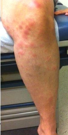
A 33-year-old woman with an unremarkable past medical history presented in clinic with complaints of frontotemporal headaches and neck pain without stiffness of 1 week’s duration. Tension headache was initially diagnosed. Four days later, she presented to the local emergency department (ED) with a worsening headache now accompanied by nausea, vomiting, ankle pain with swelling, and fevers (temperatures reported as high as 40°C [105°F]). She had a new rash on her lower extremities. The rash appeared as erythematous, tender patches, 3 to 4 cm in size, on the anterior aspect of her lower extremities bilaterally (Figure). Treatment with prednisone was started for a presumed autoimmune response. At a clinic follow-up 3 days later, her ankle pain and swelling had improved but the headaches and fevers had not relented. A biopsy of the rash was significant for superficial perivascular dermatitis with mild focal spongiosus. Because she was afebrile at the visit, the patient was instructed to continue the current dose of prednisone and seek medical attention should either fevers or headache return.
Several days later, she returned to the ED with new-onset diffuse chest pain, abdominal pain, and worsening headache. Analysis of cerebral spinal fluid (CSF) revealed leukocytosis (tube 1: WBC 51 [neutrophiils, 1%; lymphocytes, 93%; monocytes, 6%]; RBC 716; tube 4: WBC, 38 [neutrophils, 2%; lymphocytes, 88%; monocytes, 10%]; RBC, 42; glucose, 49 mg/dL; protein, 38 mg/dL) and she was admitted to the hospital for presumed meningitis. Her headaches continued during the admission but she remained afebrile. Laboratory findings at this time were significant for elevated high-sensitivity C-reactive protein (hs-CRP) (90 mg/L; normal, <10 mg/L). Results of a chest radiograph and CT scan of the head were normal. Infectious mononucleosis was ruled out (negative heterophile antibody test). Syphilis also was ruled out (RPR negative) and no evidence was found of bacteremia or infectious meningitis. She was discharged from the hospital and was referred for follow-up with rheumatology.
At the appointment with rheumatology she reported persistent low-grade fevers and recurrent arthralgias, as well as new-onset right periorbital edema, erythema, and photophobia. Profiles were negative for antinuclear antibodies, angiotensin-converting enzyme (ACE), cryoglobulin, and vasculitis. However, she had positive Lyme titers and a CT scan of the chest revealed pulmonary nodules. She was referred to ophthalmology for a comprehensive eye exam that was notable for bilateral iritis; she was given oral prednisone and doxycycline.
The patient returned to the ED 2 months later with persistent headaches, which she said were made worse by exertion and light. She also reported new joint pain, neck stiffness, and worsening fatigue. CSF leukocytosis had worsened (tube 1: WBC, 67 [neutrophils, 8%; lymphocytes, 74%; monocytes, 15%; eosinophils, 3%]; RBC, 974; tube 4: WBC, 49 [neutrophils, 5%, lymphocytes 80%, monocytes, 12%, eosinophils 3%]; glucose, 37 mg/dL; protein, 87 mg/dL) and she was readmitted to the hospital for treatment of meningitis. CSF cultures and virus studies were negative. Blood cultures, HIV antibodies, a hepatic panel, and complete blood count were normal. Antibody titers were negative for lupus erythematosus, rheumatoid factor, and ANCA. Her ESR was 14 mm/h and hs-CRP level was 10.4 mg/L. MRI study of the brain was normal. Repeated CT scan of the chest was significant for pulmonary nodules. Biopsy of lung tissue was significant for granulomatous disease and a diagnosis of Löfgren syndrome was made.
She was treated with prednisone 40 mg daily for 1 week, and her symptoms improved. She was discharged and instructed to follow up with rheumatology, pulmonology, and neurology. Prednisone was continued and tapered slowly because symptoms returned when it was discontinued. At a follow-up, she reported improvement in her headaches and arthralgias and complete resolution of erythema nodosum.
Discussion
Löfgren syndrome is a form of acute sarcoidosis characterized by a triad of symptoms: hilar adenopathy, erythema nodosum, and arthralgias.1 High-grade fevers, as seen in this patient, have been reported only rarely.2-4 Ophthalmologic involvement, also seen in our patient, is a common finding.1,3 Fifty percent of patients with Löfgren syndrome may have normal ACE levels.1,2
Sarcoidosis most commonly affects women and between the ages of 20 and 50 years.1 Only 10% of patients with sarcoidosis present with Löfgren syndrome.5 Patients with Löfgren syndrome are most commonly of Caucasian descent. Presentation of the condition varies among ethnicities. Arthralgia is a common symptom among Caucasians, and infrequent among Japanese patients.6 Swedish women with Löfgren syndrome are more likely to have erythema nodosum, but Swedish men were more likely to have periarticular inflammation of the ankles.7
Our patient presented with rarely seen high fevers and also headache. The latter is seen in patients with neurosarcoidosis, but is not typical of Löfgren syndrome.8 Also of interest, our patient was of Middle Eastern descent, a nationality in which Löfgren syndrome is rarely reported.
Löfgren syndrome is self-limited, but when symptoms are severe, therapy with corticosteroids is recommended. Löfgren syndrome spontaneously resolves over 2 years in 80% of patients.
Acknowledgments
The authors would like to thank Ms Susan Ebbinghouse for assistance in the literature search.
References:
1. Drake W, Newman L. Sarcoidosis. In: Mason RJ, Broaddus VC, Martin TR, et al, eds. Murray and Nadel's Textbook of Respiratory Medicine. 5th ed. Philadelphia: Saunders Elsevier; 2010:1431.
2. Patel KN, Patel F, Hudgins L. Löfgren’s syndrome presenting as a case of fever of unknown origin. Tenn Med. 2007;100:37-38.
3. Oshima M, Maeda H, Furonaka O, et al. Sarcoidosis with multiple organ involvement emerging as Löfgren’s syndrome. Intern Med. 2003;42:534-537.
4. Rados J, Lipozencic J, Celic D, Loncaric D. Löfgren’s syndrome presenting with erythema nodosum-like eruption. Acta Dermatovenerol Croat. 2007;15:249-253.
5. Mana J, Gomez-Vaquero C, Montero A, et al. Löfgren’s syndrome revisited: a study of 186 patients. Am J Med. 1999;107:240-245.
6. Suzuki E, Kanno T, Ohira H. Acute-onset sarcoidosis with polyarthralgia and hilar lymphadenopathy. Mod Rheumatol. 2010;20:188-192.
7. Grunewal J, Eklund A. Sex-specific manifestations of Löfgren’s syndrome. Am J Respir Crit Care Med. 2007;175:40-44.
8. LaMantia L, Erbetta A. Headache and inflammatory disorders of the central nervous system. Neurol Sci. 2004;25:S148-S153.
