- Clinical Technology
- Adult Immunization
- Hepatology
- Pediatric Immunization
- Screening
- Psychiatry
- Allergy
- Women's Health
- Cardiology
- Pediatrics
- Dermatology
- Endocrinology
- Pain Management
- Gastroenterology
- Infectious Disease
- Obesity Medicine
- Rheumatology
- Nephrology
- Neurology
- Pulmonology
Hand Deformities and Heart Problems: A Case Report
Patients who present with congenital hand deformities in association with cardiac disorders require a detailed evaluation.
Congenital deformities of the hands are common. A small group of these deformities are associated with congenital disorders of the heart. Some of the hand-heart abnormalities are inheritable.
Patients who present in primary care offices may demonstrate some form of congenital deformities in the extremities. Some patients express concern about the possibility of their offspring inheriting such deformities, especially when they have other disorders, such as heart valve disease.
Patients who present with congenital hand deformities in association with cardiac disorders require a detailed evaluation to identify the type of deformity and possible presence of hereditary hand-heart syndromes. However, most congenital hand deformities are isolated findings that pose no additional risks or associations with serious medical disorders.
Our patient, a 45-year-old African American man, presented with heart disorders and congenital hand deformities. A diagnosis of amniotic band syndrome (ABS) was made for his hand deformities.
In this case report, we present the various types of hand deformities and inheritable heart-hand syndromes as well as our patient’s deformity.
Background
In United States, some form of malformation of the upper extremities affects about 0.16% to 0.18% of the population.1 Malformations are classified into 7 types on the basis of embryogenesis (Table 1).2 A very small fraction of patients present with specific upper extremity deformities that have associated heart disorders and are hereditary (eg, about 0.95 cases per 100,000 total births present with Holt-Oram syndrome3). Heart-hand syndromes include Holt-Oram syndrome,4 Tabatznik syndrome,5 familial brachydactyly,6 and polydactyly7 (Table 2).
Table 1. Classification of Hand Malformationsa
Type
Description
I
Failure of formation
II
Failure of differentiation
III
Duplication
IV
Overgrowth
V
Undergrowth
VI
Constriction band syndromes
VII
Generalized anomalies and syndromes
a American Society for Surgery of the Hand and International Federation of Societies for Surgery of the Hand.
Table 2. Heart-Hand Syndromes
Syndrome
Description
Holt-Oram syndrome
Thumb anomalies; abnormal carpal bones and radius, with atrial septal defect; autosomal dominant
Tabatznik syndrome
Brachydactyly; telephalangy; bifid thumbs; and bowing of the radius, with arrhythmias; autosomal or X-linked trait
Familial brachydactyly
Brachydactyly of middle or proximal phalanx of digit 2 - 5, with sick sinus syndrome; autosomal dominant
Polydactyly
Polydactyly; cutaneous syndactyly, with patent ductus arteriosus; ventricular septal defect; and genital defects (hydrometrocolpos); autosomal recessive
ABS includes a group of disorders that are characterized by entrapment of various parts of the fetus by constriction bands of the amniotic membrane, acrosyndactyly, and amputations. In 300 BCE, Hippocrates postulated that external compression by the ruptured amniotic tissue results in ABS. Various other theories have been proposed, but this theory has been widely accepted as the main cause.8
Case Report
A 45-year-old African American man presented in our primary care office for evaluation and management of his medical conditions. He was born with digital abnormalities that included poor development and an absence of fingers that required surgical correction at an early age. From childhood through adulthood, he received diagnoses of hypertrophic obstructive cardiomyopathy, mitral regurgitation that required mitral valve replacement at age 4, complete heart block assisted with permanent pacemaker placement, coronary artery disease, type 2 diabetes mellitus, and hypertension.
The patient inquired about the chances of inheritance of his hand deformities in his children to find out before he and his spouse could plan for a child. The patient had no siblings. His mother did not have skeletal deformities, and she had passed away because of a “cocaine-induced heart attack." He had never met his father. He was not sure about the hand deformities in other family relatives.

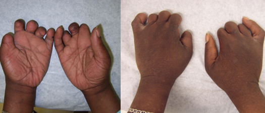
Figure 1. The appearance of the patient’s hands is seen in dorsal (left) and ventral (right) views.
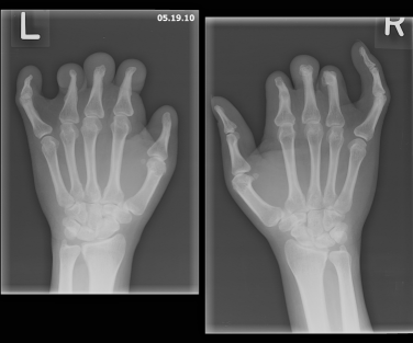
The patient was of short stature and had bilateral hand deformities in the form of extremely short nubbin-like second, third, and fourth fingers along with somewhat deformed thumbs. The fifth fingers of both hands demonstrated clinodactyly with partially hypoplastic nails (Figure 1). Radiographic images of both hands showed an absence of the middle and distal second, third, and fourth phalanges with tapering proximal phalanges and tapering of the distal fifth phalanx and distal first phalanx (Figure 2).

Figure 2. Radiographic images of the patient’s hands showed an absence of the middle and distal second, third, and fourth phalanges with tapering proximal phalanges and tapering of the distal fifth phalanx and distal first phalanx.
The patient had a good peripheral arterial pulse volume in all extremities. A detailed phenotypic analysis and chromosomal analysis (including a negative test result for TBX5 gene mutation) did not support any of the hereditary hand-heart syndromes. His hand deformities were compatible with a clinical diagnosis of ABS.
Discussion
ABS has been given many names, including ADAM complex, pseudoainhum, and congenital constriction bands. During intrauterine fetal development, strands of amniotic membrane are formed as a result of spontaneous rupture of the amniotic membrane. These strands encircle parts of the developing fetus in the form of annular constriction bands. The fetus grows, but the bands do not, leading to severe neurovascular compromise and development of various malformations of the affected part.
ABS has been reported to occur in 1 in 3000 pregnancies in the United States.9 The disorder appears to be sporadic and has no gender preference; it has been linked as a cause of about 178 miscarriages per 10,000 pregnancies.
Various causes have been proposed. Prenatal risk factors include low birth weight, premature birth, maternal exposure to certain drugs during pregnancy (eg, mifepristone, cocaine), and maternal illness during pregnancy. Possible mechanisms include a defect in the germplasm that leads to sloughening of the soft tissue,10 a lack of development of the mesoderm,11 and fetal hypoperfusion.
Although a genetic predisposition has not been found with ABS, a familial autosomal trait has been described as multiple benign circumferential skin creases. ABS has been described as a consequence of a deformation sequence characterized by the absence of an intrinsic embryonic defect rather an abnormal external structural mechanism.
The events that lead to damage caused by the encircling bands of amniotic tissue usually take place within the first 26 days of the postconception period. During early gestation, such bands may result in spontaneous abortion. In the later stages, in utero bands may wrap around extremities or digits, resulting in amputations, syndactyly, or clubfeet.
Likewise, involvement of the face, chest, and trunk results in cleft lip or palate and craniofacial abnormalities and deformities of the chest and abdomen, respectively. One or more deformities may coexist.
The most common clinical presentation includes amputation of the extremities or digits, constriction bands, and acrosyndactyly. Hand abnormalities alone constitute about 80% to 90% of ABS cases.12 Facial abnormalities, such as cleft palate and cleft lip, are seen in 50% of cases. Less common presentations include encephalocele, hemihypertrophy, and length discrepancies in the lower extremities.13,14
Deformities have been classified as the following 4 types11:
• Type 1: Simple ring constriction.
• Type 2: Ring constriction and fusion of distal bony parts, possibly with lymphedema.
• Type 3: Ring constriction with fusion of soft tissue.
• Type 4: Intrauterine amputation.
During the prenatal period, obstetric ultrasonography is the only way to detect ABS. In select cases, an elevated blood level of α-fetoprotein with normal acetylcholinesterase activity indicates associated anencephaly.
Surgical excisions of the constriction band followed by plastic reconstruction by Z-, V-Y, or W-plasty is the mainstay of treatment.15 Stabilizing the child medically before contemplation of such a procedure is important. The outcome of surgical intervention depends on the neurovascular status of the affected area. Hence, plastic reconstructions are performed in stages to avoid interruption to the vascularity of the affected area. Excision usually is not indicated for bands that are superficial.
Outcomes with early surgical intervention are better in cases with acrosyndactyly, clubfeet, cleft lip, and cleft palate. Late reconstructive procedures and occupational therapy may offer good prehensile function in acrosyndactyly. Isolated superficial bands in the extremities carry a good prognosis. Medical therapy is limited. Avoidance of potential drugs that are associated with rupture of the amniotic membranes (eg, mifepristone, cocaine) is strongly recommended.
We discussed the diagnosis of ABS with our patient, and we alleviated his concern by stating that it was not an inheritable disorder.
Conclusion
Patients with congenital deformities of the hand and coexisting cardiac conditions should be evaluated for possible inheritable heart-hand syndromes and for other noninheritable types of limb deformities. Although ABS is corrected in the early stages of life, patients with ABS should be monitored at regular intervals to assess for recurrence of constriction bands resulting from secondary contractures.
References
1. Lamb DW, Wynne-Davies R, Soto L. An estimate of the population frequency of congenital malformations of the upper limb. J Hand Surg. 1982;7A:557-562.
2. Swanson AB, Swanson GD, Tada K. A classification for congenital limb malformation. J Hand Surg. 1983;8A(5, pt 2):693-702.
3. Elek C, Vitz M, Czeizel E. Holt-Oram Syndroma. Orv Hetil. 1991;132:73-78.
4. Holt M, Oram S. Familial heart disease with skeletal malformations. Br Heart J. 1960;22:236-242.
5. Temtamy S, McKusick VA. Heart-hand syndrome II (Tabatznik syndrome). BDOAS XIV. 1978;3:241.
6. Ruez de la Fuente S, Prieto F. 3-7. Heart-hand syndrome, part III; a new syndrome in three generations. Hum Genet. 1980;55:43-47.
7. Slavotinek AM, Biesecker LG. Phenotypic overlap of McKusick-Kaufman syndrome with bardet-biedl syndrome: a literature review. Am J Med Genet. 2000;95:208-215.
8. Torpin R. Amniochorionic mesoblastic fibrous strings and amnionic bands: associated constricting fetal malformations or fetal death. Am J Obstet Gynecol. 1965;91:65-75.
9. Do TT. Streeter Dysplasia. eMedicine-Medscape Drugs, Diseases & Procedures. 2012. Available at: http://emedicine.medscape.com/article/1260337-overview. Accessed January 9, 2013.
10. Streeter G. Focal deficiencies in fetal tissues and their relation to intrauterine amputations. Contributions Embroyol Carnegie Inst. 1930;22:1-46.
11. Patterson TJ. Congenital ring-constrictions. Br J Plast Surg. 1961;14:1-31.
12. Light TR. Growth and Development of the Hand. In: Reconstruction of the Child’s Hand. Carter PR, ed. Philadelphia: Lea & Febiger; 1991:122.
13. Tanguy AF, Dalens BJ, Boisgard S. Congenital constricting band with pseudarthrosis of the tibia and fibula: a case report. J Bone Joint Surg. 1995;77A:1251-1254.
14. Zych GA, Ballard A. Constriction band causing pseudarthrosis and impending gangrene of the leg: a case report with successful treatment. J Bone Joint Surg. 1983;65A:410-412.
15. Upton J, Tan C. Correction of constriction rings. Am J Hand Surg. 1991;16:947-953.
