- Clinical Technology
- Adult Immunization
- Hepatology
- Pediatric Immunization
- Screening
- Psychiatry
- Allergy
- Women's Health
- Cardiology
- Pediatrics
- Dermatology
- Endocrinology
- Pain Management
- Gastroenterology
- Infectious Disease
- Obesity Medicine
- Rheumatology
- Nephrology
- Neurology
- Pulmonology
Giant Bullous Emphysema
A 54-year-old man presented to the ED with palpitations identified as atrial flutter and RVR. Medical history included stage IV renal-cell carcinoma, end-stage COPD, NYHA class IV heart failure, and recent pulmonary embolism. A CT scan of the thorax was ordered.
A 54-year-old man presented to the emergency department with sudden onset of palpitations. His medical history included stage IV renal cell carcinoma, end-stage COPD, NYHA class IV heart failure, recent pulmonary embolism, and ongoing tobacco abuse. Cardiac examination revealed atrial flutter and a rapid ventricular rate. The patient was given a diltiazem infusion, which restored normal sinus rhythm within 24 hours.

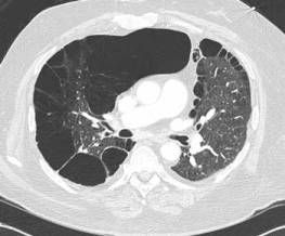
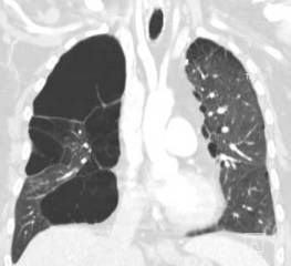
A CT scan of the thorax was ordered to help determine whether a recurrent pulmonary embolism may have contributed to the arrhythmia (Figures).
The patient also had a more than 20-year history of giant bullous emphysema (GBE). He had undergone a left-sided bullectomy in his 40s to help alleviate symptoms. He has continued to smoke and the disease has progressed.
GBE, or “vanishing lung syndrome,” is a rare condition typically found in young men who smoke. It is caused by paraseptal emphysematous bullae that coalesce and eventually compress the lung parenchyma.1 By definition, the bullae must occupy at least one-third of the hemithorax and thus can be mistaken for a pneumothorax on a standard chest film.2

Bullectomy is the preferred treatment in patients with GBE. Ideal candidates have large bullae that can be resected without sacrificing precious, functioning lung parenchyma. Localized disease allows the removal of large bullae that do not participate in gas exchange and impair ventilation. Indications include3:
• Large bullae that cause worsening dyspnea, recurrent infections, or pneumothorax.
• Bullae that occur as a manifestation of a lung carcinoma.
Results of bullectomy include increased elastic recoil of the residual lung parenchyma as well as improved mechanics of respiration. Dyspnea, exercise capacity, FEV1, and quality of life improve significantly in the first 5 years after surgery, but continue to decline thereafter.4,5 Surgery does not affect the natural history of the disease, and there are no studies that demonstrate that surgery reduces mortality.
The patient was interested in a right-sided bullectomy to ameliorate his current symptoms. He was told that his continued tobacco abuse, low cardiopulmonary reserve, and advanced malignancy made him a poor candidate. His medication regimen, which included Lopressor (metoprolol), Advair (fluticasone propionate/salmeterol), and Sutent (sunitinib malate), was not changed.
References:
1. Stern EJ, Webb WR, Weinacker A, et al. Idiopathic giant bullous emphysema (vanishing lung syndrome): imaging findings in nine patients. AJR. 1994;162:279–282.
2. Roberts L, Putman CE, Chen JTT, et al. Vanishing lung syndrome: upper lobe bullous pneumopathy. Rev Interam Radiol. 1987;12:249–255.
3. Ng CSH, Shioe ADL, Wan S, et al. Giant pulmonary bulla. Can Med Assoc J. 2001;8;369-371.
4. Palla A, Desideri M, Rossi G, et al. Elective surgery for giant emphysema: a 5-year clinical and functional follow up. Chest. 2005;128:2043–2050.
5. Neviere R, Catto M, Bautin N, et al. Longitudinal changes in hyperinflation parameters and exercise capacity after giant bullous emphysema surgery. J Thorac Cardiovasc Surg. 2006;132:1203-1207.
