- Clinical Technology
- Adult Immunization
- Hepatology
- Pediatric Immunization
- Screening
- Psychiatry
- Allergy
- Women's Health
- Cardiology
- Pediatrics
- Dermatology
- Endocrinology
- Pain Management
- Gastroenterology
- Infectious Disease
- Obesity Medicine
- Rheumatology
- Nephrology
- Neurology
- Pulmonology
GI Lesions and Gut Feelings-A Photo Essay
Here: 5 Tips about Crohn disease, Pseudopolyps, GI Cancer, and other GI Maladies
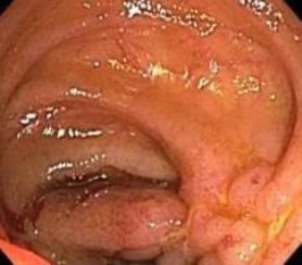
Patients with inflammatory bowel disease-especially those with Crohn disease-typically experience discomfort for 3 to 5 years before the true cause of their symptoms is identified. Symptoms are often attributed to a more common problem, such as infection or irritable bowel syndrome. Also, 20% to 40% of patients with Crohn disease or ulcerative colitis have involvement outside of the bowel that manifests as back pain, joint pain, mouth ulcerations, pyoderma gangrenosum in the legs, or erythema nodosum of the shins. These extraintestinal “red herrings” can obscure the diagnosis. This colonoscopic image shows moderate to severe inflammation of the ileum that is typical of Crohn disease.
Click here for the next image
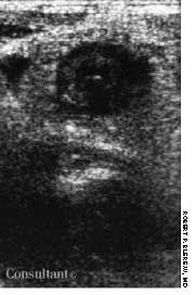
A 17-year-old man with an 18-month history of “a nervous stomach” awoke with severe pubic area pain. His temperature was 97.1°F; physical exam revealed localized tenderness at McBurney point. The WBC count was 20,300/μL, with 91% neutrophils and 9% lymphocytes. Abdominal ultrasonography demonstrated a bull’s eye typical of acute appendicitis. At surgery for anticipated appendectomy, the distal 8 to 10 inches of ileum were acutely inflamed and sausage-shaped. Approximately 1 inch of ileum adhered to the anterior abdominal wall at the suprapubic area-the apparent cause of the pubic area pain. The appendix was retrocecal and normal. An appendectomy was performed, and the patient was treated methylprednisolone.The bull’s eye sign on abdominal ultrasonography strongly suggests acute appendicitis and terminal ileitis (Crohn disease). An unusually large normal appendix also may present with the same picture.
Click here for the next image
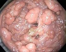
A 26-year-old man presented to the emergency department with progressively worsening generalized fatigue and weakness that had been present for the past few months. A family history was notable for colon cancer in his father and grandfather. Results of laboratory studies were significant for iron deficiency anemia. Colonoscopy revealed innumerable polyps carpeting the mucosa from the rectum to the cecum. The diagnosis: familial adenomatous polyposis.
Click here for the next image
For 6 weeks, a 29-year-old previously healthy man had between 10 and 15 episodes daily of small-volume bloody diarrhea with intermittent paraumbilical pain. Colonoscopy revealed diffuse ulceration with loss of vascularity and mucosal surfaces that extended from the rectum to the cecum. Pseudopolyps-distinct, irregular, raised areas of normal-appearing mucosa-were noted among the areas of friability, fibrous stranding, and ulceration. Pseudopolyps, which represent a combination of reactive hyperplasia and mucosal ulceration, are not uncommon in severe or chronic ulcerative colitis.
Click here for the next image
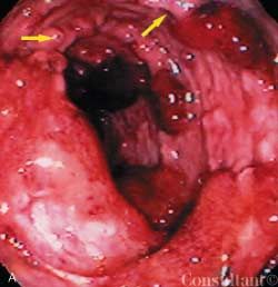
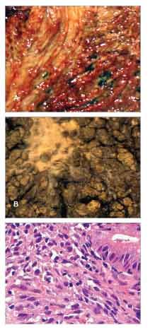
Kaposi sarcoma (KS) commonly involves the GI tract in patients who have HIV/AIDS. Patients with GI KS are typically asymptomatic, although gastric outlet obstruction and bleeding have been described. The gross appearances include macular, erythematous maculopapular, and polypoid lesions.
Click here to return to the first image.
Clinical Tips for Using Antibiotics and Corticosteroids in IBD
January 5th 2013The goals of therapy for patients with inflammatory bowel disorder include inducing and maintaining a steroid-free remission, preventing and treating the complications of the disease, minimizing treatment toxicity, achieving mucosal healing, and enhancing quality of life.
