- Clinical Technology
- Adult Immunization
- Hepatology
- Pediatric Immunization
- Screening
- Psychiatry
- Allergy
- Women's Health
- Cardiology
- Pediatrics
- Dermatology
- Endocrinology
- Pain Management
- Gastroenterology
- Infectious Disease
- Obesity Medicine
- Rheumatology
- Nephrology
- Neurology
- Pulmonology
Gastrointestinal Involvement of Systemic Amyloidosis
The authors present a case of AL amyloidosis with rare GI involvement and an equally rare presenting symptom.
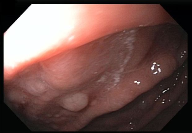
Figure 1. Nodules seen on EGD.

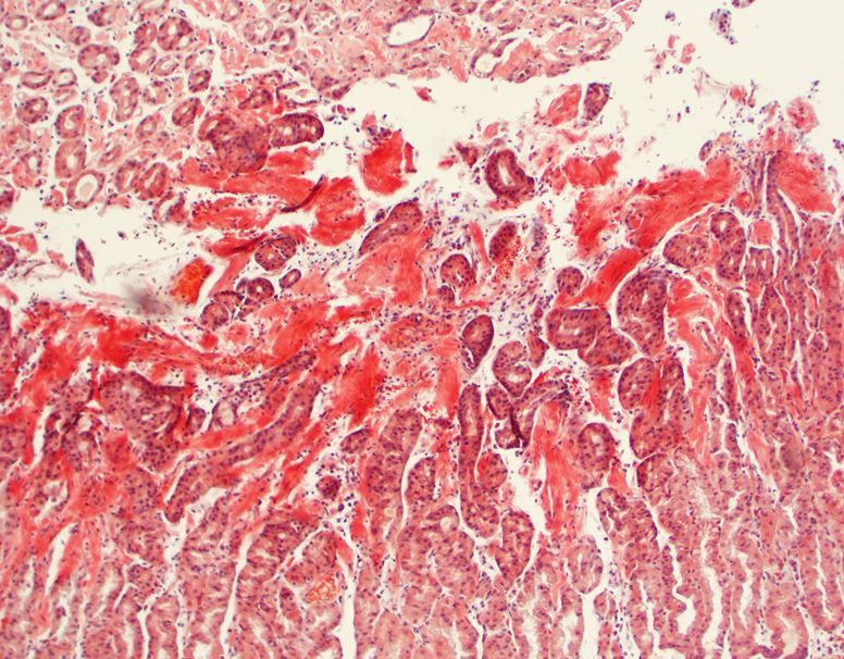
Figure 2. Biopsied tissue from nodules.

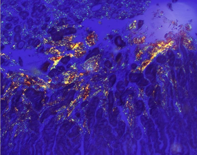
Figure 3. AL amyloid deposits.

Amyloidosis is a rare disease of abnormal protein folding, which often presents with multiple organ involvement. The most common organs affected are the heart and the kidneys. Involvement of the gastrointestinal (GI) system is rare, occurring in about 3% of cases.1 When GI system involvement occurs, presentation may include signs and symptoms of GI bleeding. We present a patient who had symptoms of upper GI bleeding which lead to esophagogastroduodenoscopy (EGD) with biopsy which was diagnostic for amyloidosis.
Case presentation
A 72-year-old man with a history of peptic ulcer disease presented to the emergency department (ED) with subacute dark tarry stools, and progressive fatigue. A CBC revealed mild anemia (13.4 g/dL) and fecal occult blood testing was positive. The patient was admitted for possible upper GI bleeding. An esophagogastroduodenoscopy (EGD) was performed. It revealed mild erythema with nodularity of the fundus and body of the stomach (Figure 1; please click on all figures to enlarge). No active bleeding, gastric or esophageal varicies, or ulcers were seen. Biopsies were obtained from the nodules (Figure 2). Gastric biopsies revealed amyloid deposits, positive for Congo red stain with characteristic apple green birefringence under polarized light, indicative of AL amyloid deposits (Figure 3).
Before biopsy results were known, additional history revealed progressive lower extremity edema and increasing abdominal girth of several months’ duration. Additional laboratory evaluation revealed elevations of AST 111 IU/L (5.0 - 34 IU/L) and alkaline phosphatase 645 IU/L (40 - 150 U/L) and INR 1.3 as well as reduced levels of serum albumin 1.5 g/dL (3.5 - 5.0 g/dL), suggestive of liver dysfunction. An abdominal ultrasound showed ascites with a normal appearing liver and normal size spleen. Paracentesis was performed and ascitic fluid analysis revealed a SAAG of 1.5 and protein of 0.8 g/dL, suggestive of portal hypertension. A transjugular liver biopsy was performed for prognostic purposes; it revealed a complete replacement of hepatic parenchyma with amyloid deposition.
Other organ systems also showed dysfunction including the heart (troponin 0.383 ng/mL, B-natriuretic peptide 3014 pg/ml) in which a 2D echocardiogram revealed left ventricular hypertrophy with diastolic dysfunction and preserved ejection fraction. Also, a creatinine level of 1.6 mg/dL and nephrotic-range proteinuria (>500mg/dL) are indicative of nephrotic syndrome.
The constellation of findings placed the patient category of “very high risk” disease, with a median survival of 3-6 months.2 After discussion of available options, the patient elected to be discharged with home hospice. For alleviation of his orthostatic symptoms, the patient was discharged on midodrine 50mg PO TID, which he reported had improved his symptoms during his inpatient stay. Many drugs typically used in cardiac failure (beta blockers, ACE inhibitors, etc.) are ineffective and sometimes contraindicated in patients with amyloid heart disease.3
Discussion
Amyloidosis is a systemic disease that may present with a constellation of symptoms. When present in the liver, amyloid deposits may result in portal hypertension and liver dysfunction. GI tract involvement occurs infrequently, but when present 45% to 75% of affected patients will have occult bleeding.
In this case, the EGD and gastric biopsy suggest occult gastric bleeding due to amyloid deposits compromising the integrity of the stomach mucosal wall.
Conclusion
Amyloidosis is a systemic disease that may involve multiple organs. GI tract involvement occurs infrequently, but when present, affected patients will likely have occult bleeding. This may be from ulceration, erosions, or without specific source. This is a rare case in which a presenting symptom of amyloidosis was GI bleeding and diagnosis was first made by endoscopic biopsy.
References:
1. Cowan AJ, Skinner M, Seldin DC, et al. Amyloidosis of the gastrointestinal tract: a 13-year, single-center, referral experience. Haematologica. 2013;98:141-146. doi:10.3324/haematol.2012.068155.
2. Kastritis E, Dimopoulos MA. Recent advances in the management of AL Amyloidosis. Br J Haematol. 2015;22. doi: 10.1111/bjh.13805.
3. Desport E, Bridoux F. Al amyloidosis. Orphanet J Rare Dis. 2012 Aug 21;7:54. doi: 10.1186/1750-1172-7-54.
Clinical Tips for Using Antibiotics and Corticosteroids in IBD
January 5th 2013The goals of therapy for patients with inflammatory bowel disorder include inducing and maintaining a steroid-free remission, preventing and treating the complications of the disease, minimizing treatment toxicity, achieving mucosal healing, and enhancing quality of life.
