- Clinical Technology
- Adult Immunization
- Hepatology
- Pediatric Immunization
- Screening
- Psychiatry
- Allergy
- Women's Health
- Cardiology
- Pediatrics
- Dermatology
- Endocrinology
- Pain Management
- Gastroenterology
- Infectious Disease
- Obesity Medicine
- Rheumatology
- Nephrology
- Neurology
- Pulmonology
Foreign-Body Retrieval: Diamonds in the Rugae
Progressive weakness, confusion, and decreased oral intake preceded hospital admission for this 73-year-old man with a history of Parkinson dementia and resection for esophageal adenocarcinoma. The real problem, seen here, was revealed on a chest x-ray film.
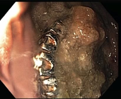
A 73-year-old man with a medical history significant for Parkinson dementia and esophageal adenocarcinoma with surgical resection and anastomosis at the gastroesophageal junction (GEJ) was admitted to the hospital following 3 weeks of progressive weakness, confusion, and decreased oral intake. Most of the patient’s symptoms were attributed to a urinary tract infection; however, a chest x-ray film revealed 3 peculiar ring-like structures within the stomach (Figure 1). The patient subsequently underwent an esophagogastroduodenoscopy (EGD) that revealed a large amount of food debris in the stomach in addition to what appeared to be a large diamond ring flanked by 2 gemstone-studded bands (Figure 2). En bloc retrieval of the entire treasure was initially attempted with a Roth® net; however, the size of the objects prohibited removal past the post-surgical anastomosis at the GEJ.


Figure 1
Figure 2

Figure 3
The use of an overtube was also restricted by the post-surgical anatomy at the site. The rings were placed back into the stomach and removed individually using a grasping device. The entire ring set was successfully removed (Figure 3). (Value was estimated to be more than $11,000.)
The patient’s wife explained that the rings had been missing for more than 3 months. Apparently the patient had inadvertently ingested the jewelry. Prolonged retention of the rings was most likely the result of post-surgical delayed gastric emptying. The patient did well following the procedure and was discharged home in stable condition. His wife was warned about leaving such small items available to her husband.
Discussion
While cases of accidental ingestion occur more frequently in the pediatric population, adults with medical histories compounded by substance abuse or neuropsychiatric disorders are also at risk.1 In our case, the patient’s somewhat poorly defined clinical features of confusion and decreased oral intake easily might have been attributed to his urinary tract infection or progressive cognitive disease. Only with routine imaging was a more insidious culprit discovered.
In approximately 90% of patients who have ingested a foreign body, the object will pass without requiring intervention.2 The use of endoscopic and/or surgical retrieval is often necessary when the presence of a foreign body leads to airway compromise, potential impaction or, as in our patient’s case, worsening mucosal damage with prolonged retention.2 Reports indicate that approximately 9% of cases requiring retrieval can be complicated by other pathology, such as strictures and mass lesions.3 Our patient’s surgical history provides a case in point.
His esophagectomy, while ultimately lifesaving, led to post-vagotomy gastroparesis and a significant period of retention. A study by Sutcliffe et al4 found that this type of complication affects up to 12% of patients following esophagectomy. The narrowed post-surgical area at the GEJ also proved to be the limiting factor in both the removal of the foreign objects and employment of an overtube-a device often used to help protect the mucosa during such procedures.5
In a case of foreign-body ingestion, determine first that there is no imminent threat to the patient. Should endoscopy be warranted, one should be prepared to tailor the retrieval technique to the nature of the objects and to unforeseen anatomic or pathologic variants.
References1. ASGE Standards of Practice Committee; Ikenberry SO, Jue TL, Anderson MA, et al. Management of ingested foreign bodies and food impactions. Gastrointest Endosc. 2011;73:1085-1091.
2. Li ZS, Sun ZX, Zou DW, et al. Endoscopic management of foreign bodies in the upper-GI tract: experience with 1088 cases in China. Gastrointest Endosc. 2006;64:485-492.
3. Zhang S, Cui Y, Gong X, et al. Endoscopic management of foreign bodies in the upper gastrointestinal tract in South China: a retrospective study of 561 cases. Dig Dis Sci. 2010;55:1305-1312.
4. Sutcliffe RP, Forshaw MJ, Tandon R, et al. Anastomotic strictures and delayed gastric emptying after esophagectomy: incidence, risk factors and management. Dis Esophagus. 2008;21:712-717.
5. Tierney W, Adler D, Conway J, et al. Overtube use in gastronintestinal endoscopy. Gastrointest Endosc. 2009;70:828-834.
Clinical Tips for Using Antibiotics and Corticosteroids in IBD
January 5th 2013The goals of therapy for patients with inflammatory bowel disorder include inducing and maintaining a steroid-free remission, preventing and treating the complications of the disease, minimizing treatment toxicity, achieving mucosal healing, and enhancing quality of life.
