- Clinical Technology
- Adult Immunization
- Hepatology
- Pediatric Immunization
- Screening
- Psychiatry
- Allergy
- Women's Health
- Cardiology
- Pediatrics
- Dermatology
- Endocrinology
- Pain Management
- Gastroenterology
- Infectious Disease
- Obesity Medicine
- Rheumatology
- Nephrology
- Neurology
- Pulmonology
Fordyce Angiokeratoma
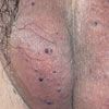
A 52-year-old man presented with asymptomatic papules on his scrotum. The lesions had first appeared 1 year earlier. He had not sustained local trauma to the scrotum, and his medical history was unremarkable. There was no family history of similar skin lesions.
A 52-year-old man presented with asymptomatic papules on his scrotum. The lesions had first appeared 1 year earlier. He had not sustained local trauma to the scrotum, and his medical history was unremarkable. There was no family history of similar skin lesions.

Numerous dark purple, dome-shaped papules were scattered over the scrotum. They varied in diameter from 2 to 4 mm. The physical findings were otherwise normal. Specifically, the patient had no associated varicocele or inguinal hernia. The clinical diagnosis was Fordyce angiokeratoma.
ANGIOKERATOMA OF THE SCROTUM: AN OVERVIEW
Angiokeratomas are characterized by ectasia of the superficial dermal vessels and hyperkeratosis of the overlying epidermis.1The dilated blood vessels are lined by a thin layer of endothelial cells.2 A narrow zone of collagen separates the vascular ectasia from the overlying epidermis. Hyperkeratosis is typically mild or absent.2 Acanthosis varies with the age of the lesion.2
Five types of angiokeratomas are recognized3:
• Angiokeratoma of the scrotum or vulva (Fordyce type).
• Solitary or multiple papular angiokeratoma.
• Mibelli angiokeratoma.
• Angiokeratoma circumscriptum.
• Angiokeratoma corporis diffusum.
Angiokeratoma of the scrotum was first described in 1896 by Fordyce, who reported the case of a 60-year-old man who had angiokeratoma that was associated with bilateral varicoceles.4
The onset is usually after the age of 40 years.5 The prevalence is reported to increase with age, from 0.6% in 16-year-old males to 17% in those older than 70 years.5 The condition is not familial.5
Etiology. The exact cause is unknown. Some experts regard the lesion as a degenerative disorder.6 Local venous hypertension might play a causative role given that the condition is more common in patients with coexisting varicocele, hydrocele, inguinal hernia, benign prostatic hypertrophy, or hemorrhoid.7 Agger and Osmundsen8 reported regression of angiokeratomas of the scrotum in a patient after surgical treatment of a coexisting varicocele. However, Orvieto and colleagues9 found no association between varicocele and angiokeratoma of the scrotum.
Clinical features. Fordyce angiokeratoma typically affects the scrotum.10 Occasionally, the lesions may develop on the penis.11,12 An equivalent variant affects the vulva, labia majora, and clitoris in older women.13,14
Fordyce angiokeratoma classically presents as multiple dark red, dome-shaped papules of 2 to 5 mm in diameter, with a discrete keratotic surface.2,10 Lesions of recent onset are red, soft, and compressible, whereas older lesions are dark blue to purple, firm, and noncompressible.3
Complications. Angiokeratoma of the scrotum can lead to diffuse redness of the scrotum if numerous surface blood vessels become dilated.5,15 Bleeding without preceding trauma is rare.16,17 The lesions can cause the patient anxiety and embarrassment.10
Differential diagnosis. Fordyce angiokeratoma must be distinguished from angiokeratoma corporis diffusum, which is the only other angiokeratoma that can involve the genitalia. The latter is characterized by numerous tiny red papules in a symmetrical distribution over the entire body. Other conditions in the differential diagnosis are nevomelanocytic nevus and melanoma.
Management. The condition is benign, and treatment is usually unnecessary. Excision, electrodessication, or laser therapy may be considered for symptomatic lesions or for cosmesis.5,10
References:
REFERENCES:
1.
Karthikeyan K, Sethuraman G, Thappa DM. Angiokeratoma of the oral cavity and scrotum.
J Dermatol
. 2000;27:131-132.
2.
Schiller PI, Itin PH. Angiokeratomas: an update.
Dermatology.
1996;193:275-282.
3.
Jansen T, Bechara FG, Sticker M, et al. Angiokeratoma of the scrotum (Fordyce type) associated with angiokeratoma of the oral cavity.
Acta Derm Venereol
. 2002;82:208-210.
4.
Fordyce JA. Angiokeratoma of the scrotum.
J Cutan Genito Urin Dis
. 1896;14:81-87.
5.
Bechara FG, Jansen T, Wilmert M, et al. Angiokeratoma Fordyce of the glans penis: combined treatment with erbium: YAG and 523 nm KTP (frequency doubled neodynium: YAG) laser.
J Dermatol
. 2004;31:943-945.
6.
Atherton DJ, Moss C. Angiokeratoma of the scrotum. In: Burns T, Breathnach S, Cox N, et al.
Rook's Textbook of Dermatology.
Hong Kong: Blackwell; 2004:15. 89-15. 90.
7.
Erkek E, Basar MM, Bagci Y, et al. Fordyce angiokeratomas as clues to local venous hypertension.
Arch Dermatol
. 2005;141:1325-1326.
8.
Agger P, Osmundsen PE. Angiokeratoma of the scrotum (Fordyce). A case report on response to surgical treatment of varicocele.
Arch Derm Venereol
. 1970;50:221-224.
9.
Orvieto R, Alcalay J, Leibovitz I, Nehama H. Lack of association between varicocele and angiokeratoma of the scrotum (Fordyce).
Mil Med
. 1994;159:523-524.
10.
Lapidoth M, Ad-El D, David M, Azaria R. Treatment of angiokeratoma of Fordyce with pulsed dye laser.
Dermatol Surg
. 2006;32:1147-1150.
11.
Bechara FG, Huesmann M, Stücker M, et al. An exceptional localization of angiokeratoma of Fordyce on the glans penis.
Dermatolog
y. 2002;205:187-188.
12.
Pianezza ML, Singh D, van der Kwast T, Jarvi K. Rare case of recurrent angiokeratoma of Fordyce on penile shaft.
Urology
. 2006;68:891.e1-3.
13.
Haidopoulos DA, Rodolakis AJ, Elsheikh AH, et al. Vulvar angiokeratoma following radical hysterectomy and radiotherapy.
Acta Obstet Gynecol Scand.
2002;81:466-467.
14.
Yamazaki M, Hiruma M, Irie H, Ishibashi A. Angiokeratoma of the clitoris: a subtype of angiokeratoma vulvae.
J Dermatol
. 1992;19:553-555.
15.
Miller C, James WD. Angiokeratoma of Fordyce as a cause of red scrotum.
Cutis.
2002;69:50-51.
16.
Hoekx L, Wyndaele JJ. Angiokeratoma: a cause of scrotal bleeding.
Acta Urol Belg.
1998;66:27-28.
17.
Taniguchi S, Inoue A, Hamada T. Angiokeratoma of Fordyce: a cause of scrotal bleeding.
Br J Urol.
1994;73:589-590.
