- Clinical Technology
- Adult Immunization
- Hepatology
- Pediatric Immunization
- Screening
- Psychiatry
- Allergy
- Women's Health
- Cardiology
- Pediatrics
- Dermatology
- Endocrinology
- Pain Management
- Gastroenterology
- Infectious Disease
- Obesity Medicine
- Rheumatology
- Nephrology
- Neurology
- Pulmonology
Escherichia coli in Traveler's Diarrhea
Traveler's diarrhea (TD) occurs in persons traveling fromindustrialized countries to less developed regions of the world.Because of the growing ease of travel and an increasinglyglobalized economy, TD is becoming more common. Increasingantibiotic resistance among causative bacterial organisms andalso emergence of new pathogens are additional challenges inthe management of TD. Enterotoxigenic and enteroaggregativepathotypes of Escherichia coli are the principal causes of TD.This review discusses the epidemiology of these pathogens, aswell as elements of prevention, diagnosis, and management.[Infect Med. 2008;25:264-276]
Traveler's diarrhea (TD) affects approximately 30% to 40% of persons traveling from industrialized countries to less developed areas,1,2 resulting in an estimated 15 to 20 million cases of TD globally per year.3-5 Classic TD in adults is usually defined as more than 3 unformed bowel movements occurring within 24 hours, accompanied by other symptoms, such as cramps, nausea, fever, bloating, malaise, bloody stools, fecal urgency, or vomiting.4,6 In 1985, an NIH consensus panel defined TD as a syndrome characterized by a 2-fold or greater increase in the frequency of unformed bowel movements with commonly associated symptoms as noted above.1,5 This definition applies to both children and adults. Rates of frank dysentery, usually defined as invasive diarrhea with fever or diarrhea including bloody stools, may represent as many as 10% of cases.1,4
Risk factors for TD are both endogenous and exogenous. Exogenous risk factors include travel to high-risk destinations, such as the Indian subcontinent, Latin America, Africa, and the Middle East; the first 10 days of travel; longer durations of stay abroad; traveling in primitive conditions; and not following recommended dietary restrictions for safe food and water consumption. Endogenous risk factors include young age, immunosuppression, pregnancy, the presence of inflammatory bowel disease or diabetes mellitus, and the use of H2 blockers or antacids.7
TD usually begins during the first 2 weeks of travel but may occur anytime during the trip or after return.1,4 Children younger than 3 years are more likely to have prolonged illness than are adults (average, 29.5 days compared with 2.6 to 8.4 days in older children, adolescents, and adults).1,6 Of interest, the incidence of TD in 5-star hotels may be slightly higher than in 3- or 4-star hotels, possibly because food is more frequently prepared by hand in higher end hotels.5 The type of travel also may play a role in the incidence of TD. Beach vacations in resorts are associated with lower attack rates (28%) than are tours (31% to 32%), with adventure tours being associated with the highest rates (34%).4,8
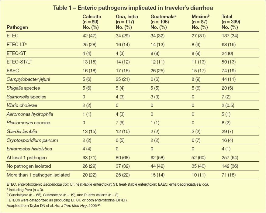
Well-designed, comprehensive epidemiological studies have shown that numerous infectious agents are implicated in TD (Table 1). The majority of cases are attributable to bacterial pathogens. Enterotoxigenic Escherichia coli (ETEC) and enteroaggregative E coli (EAEC) pathotypes combined account for approximately 50% of cases, with the remainder of cases being attributable to a large number of other bacteria, protozoa, and viruses. Of interest, even in carefully conducted studies, the cause of TD remains undetected in 10% to 40% of cases.1-3,9-11

Table 1
ENTEROTOXIGENIC E COLI
ETEC is defined as an E coli strain that produces heat-labile enterotoxin (LT) or heat-stable enterotoxin (ST).12,13 The common mechanism of pathogenesis includes colonization of the small bowel facilitated by hairlike fimbrial colonization factor antigens followed by enterotoxin secretion. LT is closely related to cholera toxin and similarly acts by raising intracellular cyclic adenosine monophosphate. ST is small and nonimmunogenic and acts by stimulating guanylate cyclase at the apical plasma membrane.
Diarrhea produced by ETEC is secretory in nature. The disease typically begins with sudden onset of watery stools lacking blood or inflammatory cells, and it is often associated with vomiting. In severe cases, the diarrhea can precipitate dehydration and electrolyte abnormalities, including hyponatremia, hypokalemia, and acidosis. Patients are usually afebrile. Almost all cases are self-limited, resolving in 3 to 4 days. With adequate attention to hydration, mortality is very low (less than 1%).14
ETEC occurs in all developing countries. It is endemic year-round but is most common during the warm and rainy seasons.14 In hospital- based studies, strains of ETEC that produce both LT and ST or ST alone were found to cause more severe disease than strains that produce LT alone.14,15
Because the diarrhea is self-limited, detection of ETEC as a cause of diarrhea is seldom necessary. No readily available test exists. Reference and research laboratories most commonly identify the organism by detection of the genes encoding LT or ST. Polymerase chain reaction (PCR) testing is very efficient for this purpose. More traditional tests include DNA probes and immunoassays for the toxins themselves.14,16 Real-time PCR is currently the most sensitive method for detecting ETEC in fecal samples.17 This test can be performed on E coli colonies isolated from a patient's stools, but it also can be performed directly on stools with excellent results. Protocols are available so that any laboratory with real-time PCR capability can perform these assays.17
ENTEROAGGREGATIVE E COLI
EAEC is defined by its unique "stacked brick" pattern of adherence to cultured human HEp-2 epithelial cells.
18
Because the HEp-2 adherence test is never performed in commercial clinical laboratories, diarrhea caused by the EAEC pathotype is substantially under diagnosed and the organism is under appreciated. Globally, the EAEC pathotype is one of the most common causes of childhood diarrhea,
19
and it is among the most common diarrheal agents in the United States.
20
Like ETEC, EAEC is noninvasive. It appears to cause diarrhea by colonization of the intestinal mucosa followed by the release of enterotoxins and cytotoxins. Cultured epithelial cells infected with EAEC exhibit greater cell damage and greater release of proinflammatory cytokines, such as interleukin (IL)-1 and IL-8, than do cultured epithelial cells infected with ETEC.18
EAEC typically elicits a mucoid, watery diarrhea that lasts for 2 to 7 days. As would be expected from in vitro pathogenesis studies, inflammation is more common in EAEC infected patients than in those with ETEC infection, but most patients are afebrile and have little or no incidence of blood in stools.19
In some patients with EAEC-associated diarrhea, persistent symptoms develop, last for up to 3 months, and may cause complications. Although both ETEC and EAEC have been implicated in postinfectious irritable bowel syndrome (IBS),21 a recent study suggests that of the two, EAEC might be particularly important as a cause of IBS.22
Several studies have suggested that EAEC may be particularly severe in immunocompromised hosts and infants. Indeed, EAEC infection may commonly occur in HIV-positive patients both in the United States and in developing countries, and the infection may be associated with severe prolonged diarrhea.19 A recent study suggested that a specific nucleotide polymorphism upstream of the IL-8 promoter may render patients more likely to become symptomatic following EAEC infection. 23 If corroborated, such data suggest that certain patients may predictably be more likely to experience TD when traveling to developing countries. This may be a particularly important area of future study, and it could have significance in managing prevention of infection.
Like ETEC, EAEC can be detected by identification of E coli carrying pathotype-specific virulence factors. Real-time PCR analysis is very sensitive for detecting these targets directly in stool,17 whereas standard block PCR testing provides adequate sensitivity once isolated E coli colonies are purified from the stool. Testing 3 colonies per fecal sample is generally sufficient. However, unlike ETEC, the precise gene targets for EAEC identification are not known with certainty. Studies have typically used the aatA gene (formerly known as CVD432), but many strains of EAEC harboring aatA are probably not human pathogens. Perhaps multiple genetic targets (such as both aatA and aaiC) must be detected to identify a true EAEC pathogen.18 In lieu of definitive data in this regard, however, the authors recommend use of the aatA gene as the most convenient diagnostic target.20
DIFFERENTIAL DIAGNOSIS
Because most cases of TD are caused by bacterial pathogens, the differential diagnosis should involve distinguishing among the bacterial agents.
Shigella, Salmonella, and Campylobacter
24
infection may each cause acute watery diarrhea, but the infection is more likely to result in signs and symptoms associated with inflammation, including fever; significant abdominal pain; and the presence of fecal blood, pus, and mucus. Dysentery syndrome (scant bloody stools with tenesmus and severe abdominal pain) is virtually never caused by ETEC or EAEC.
In areas where Vibrio species, including Vibrio cholerae, are endemic, particularly on the Indian subcontinent, this pathogen may cause severe cases of TD. ETEC and Vibrio species may cause similar severe watery diarrhea; in areas where these organisms are endemic, such disease should be treated with agents effective against both (such as ciprofloxacin). Mild watery diarrhea may be caused by viral agents or by EAEC or ETEC. Diarrhea caused by any of these organisms may remit spontaneously within 3 days, but watery diarrhea persisting for longer periods should prompt suspicion of ETEC or EAEC. Protozoal parasites (eg, Giardia, Cryptosporidium, Entamoeba histolytica, Cyclospora) may occasionally cause TD, and these should be suspected when diarrhea persists despite antimicrobial therapy.
PREVENTION
Prevention of TD begins with attention to fecal-oral hygiene. Food should be consumed cooked and still warm (preferably steaming); peeled fruits are also generally safe when peeled by the consumer. Hot beverages are safe, as are those from bottles and cans. Use of prophylactic antibiotics is discouraged, but many experts suggest that travelers have an antibiotic on hand and recommend that it be taken at the start of diarrhea (typically after 2 or 3 episodes of passing watery stools). No vaccine is available in the United States to prevent any of the common causes of TD. Use of any vaccine marketed for TD should be accompanied by the caveat that protection is partial and against only 1 of the many potential pathogens. Therefore, vaccines may confer a greater risk from false security than the marginal benefit accrued.
Although prophylactic antimicrobial therapy has been shown to be highly effective in preventing infection caused by both ETEC and other bacteria,25 considerable concern persists regarding widespread prophylactic use of antibiotics. First, there is concern that a patient might experience an adverse reaction without access to medical care. Second, indiscriminate antibiotic use increases the pressure toward development of antibiotic resistance among TD pathogens. Expert guidelines discourage routine use of antibiotic prophylaxis for TD.5,25 Some immunocompromised patients may need prophylactic antibiotic therapy, but these persons should consult physicians who are familiar with their specific conditions.
MANAGEMENT
Antibiotic therapy
Management of TD must begin with attention to hydration. Travelers should be counseled regarding the signs of dehydration and the need to consume adequate amounts of clear liquids with appropriate electrolyte content. Carrying oral rehydration salt packets may be wise when traveling to areas with endemic cholera or to remote sites. Empirical antibiotic regimens are a very effective approach to therapy.
26
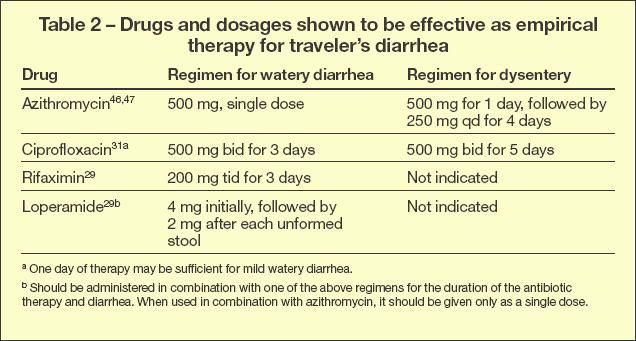
Table 2 lists recommended agents and dosages. Cases of acute nondysenteric TD are most likely caused by ETEC and EAEC, and empirical therapy should be directed toward these agents. Generally, fluoroquinolones are the first-line oral medications for treatment of dysentery.

Table 2
Campylobacter jejuni infection is becoming a worldwide problem, particularly where poultry products and antibiotics are used in animal feed. Rates of fluoroquinolone resistance among Campylobacter isolates have risen in recent years, and reduced fluoroquinolone efficacy has been observed.24,27 A recent study has shown that single-dose azithromycin is superior for empirical therapy for TD acquired in Thailand, where the highest rate of C jejuni resistance has been found, and is a reasonable first-line option for empirical management.6,27 Trimethoprim/ sulfamethoxazole and doxycycline are no longer recommended because of resistance.
The returning traveler with persistent diarrhea is more likely to have an EAEC, Shigella, Campylobacter, or parasitic infection. Stool culture and parasite detection should be undertaken, followed by therapy tailored to the agent. Pathogen-negative persistent diarrhea may be caused by postinfectious disaccharide deficiency or by IBS. The former can be readily diagnosed by response to a lactose-free diet.
Rifaximin is a relatively new, poorly absorbed (less than 0.4%) oral antibiotic that has been shown to be effective for the treatment of TD caused by noninvasive pathogens- principally ETEC and EAEC. In multiple studies, rifaximin was shown to be safe and well tolerated.10,24,28 Studies evaluating the response of EAEC strains to rifaximin therapy have shown similar results with improvement in duration of EAEC-associated diarrhea from 72 to 22 hours.9 In another recent study, rifaximin was effective in shortening the duration of illness and decreasing the passage of unformed stools in TD attributable to known pathogens and in pathogen-negative diarrhea without definable cause. This is consistent with the hypothesis that the most probable basis for TD without definable cause is undetected bacterial pathogens.10
TD poses special risks for both pregnant women and children, and prophylactic regimens should be adjusted accordingly. Prophylaxis for children can consist of either azithromycin (10 mg/kg on day 1 and 5 mg/kg on days 2 and 3) or ciprofloxacin (10 to 20 mg/kg given every 12 hours for 3 days). The prophylactic regimen of choice for pregnant women is azithromycin at a dosage of 500 mg/d for 3 days. Rifaximin is not approved for pregnant women or children younger than 12 years.
Nonantibiotic therapy With the emergence of resistant organisms, nonantibiotic therapeutic options for TD are garnering increased attention. The antimotility agent loperamide has been shown to reduce the passage of loose stools by about 50%; however, most experts recommend using loperamide in combination with an antibacterial agent because use alone may aggravate the infection.25,29-32 The American Academy of Pediatrics does not recommend the use of antimotility drugs in children. The probiotic bacterium Lactobacillus GG has shown some benefit in treating diarrhea in children and also may be useful prophylactically to help to reduce the incidence of diarrhea.33,34
POSTINFECTIOUS IBS
Postinfectious IBS (PI-IBS) is vaguely defined as the onset of IBS symptoms after an episode of enteric infection. 35 Compared with all IBS, PIIBS is more often characterized by diarrheal symptoms, with less frequent bouts of constipation and abdominal pain. Up to 10% of patients who have had TD will develop PIIBS, 4,36 which may persist for months or years. In a long-term (6 years) follow- up study of 192 patients treated for gastroenteritis, recovery of normal bowel function was observed in only 43% of those in whom PI-IBS symptoms developed.2,37 In another study of patients with IBS, decreased prevalence of the anti-inflammatory cytokine IL-10 and transforming growth factor was observed, implying that these patients may be more susceptible to prolonged and severe inflammation.38
THE FUTURE OF TD
Because of increasing global travel, TD is becoming a growing problem. Any effective preventive approach will need to provide protection against a wide range of serologically diverse enteric pathogens. Killed ETEC vaccines that are currently available provide only moderate efficacy, 39 predominantly against severe disease. However, the ease of manufacture and delivery of these vaccines suggest that a cocktail of multiple killed agents could theoretically be developed. Clinical trials of live, attenuated bacterial vaccines for infections attributable to ETEC, Shigella species, and V cholerae are being conducted.40 These vaccines promise greater immunogenicity than killed vaccines, but delivery of multiple microbial antigens will require sophisticated feats of molecular engineering. Fortunately, such strategies are feasible: investigators at the University of Maryland, for example, have engineered Shigella flexneri strains that express the fimbrial surface antigens of ETEC.41 Transcutaneous immunization has also been proposed and may provide another method of multiple antigen delivery.42
Nonvaccine interventions in development include probiotic agents43 and pooled immunoglobulins.44,45 A meta-analysis of studies of probiotics for prevention of TD found a pooled relative risk in favor of significant protection.43 Although this is a promising observation, larger clinical trials of a consistent probiotic preparation are needed. Orally administered immunoglobulins offer some promise,44,45 but no polyvalent preparations are in late development. Prevention of TD is currently best achieved by careful attention to fecal-oral hygiene.
References:
- Staat MA. Travelers’ diarrhea. Pediatr Infect Dis J. 1999;15:373-374.
- Connor BA. Sequelae of travelers’ diarrhea:focus on postinfectious irritable bowel syndrome. Clin Infect Dis. 2005;41(suppl 8):S577-S586.
- Guerrant RL, Oria R, Bushen OY, et al. Global impact of diarrheal diseases that are sampled by travelers: the rest of the hippopotamus. Clin Infect Dis. 2005;41(suppl 8):S524-S530.
- Steffen R. Epidemiology of traveler’s diarrhea. Clin Infect Dis. 2005;41(suppl 8):S536-S540.
- Hill DR, Ericsson CD, Pearson RD, et al. The practice of travel medicine: guidelines by the Infectious Diseases Society of America. Clin Infect Dis. 2006;43:1499-1539.
- Mackell S. Traveler’s diarrhea in the pediatric population: etiology and impact. Clin Infect Dis. 2005;41(suppl 8):S547-S552.
- Rack J, Wichmann O, Kamara B, et al. Risk and spectrum of diseases in travelers to popular tourist destinations. J Travel Med. 2005;12:248-253.
- Steffen R, Van der Linde F, Gyr K, Schär M. Epidemiology of diarrhea in travelers. JAMA. 1983;249:1176-1180.
- Infante RM, Ericsson CD, Jiang ZD, et al. Enteroaggregative Escherichia coli diarrhea in travelers: response to rifaximin therapy. Clin Gastroenterol Hepatol. 2004;2:135-138.
- DuPont HL, Haake R, Taylor DN, et al. Rifaximin treatment of pathogen-negative travelers'diarrhea. J Travel Med. 2007;14:16-19.
- Wilson ME. Diarrhea in nontravelers: risk and etiology. Clin Infect Dis. 2005;41(suppl 8): S541-S546.
- Nataro JP, Kaper JB. Diarrheagenic Escherichia coli. Clin Microbiol Rev. 1998;11:142-201.
- Kaper JB, Nataro JP, Mobley HL. Pathogenic Escherichia coli. Nat Rev Microbiol. 2004;2:123-140.
- Qadri F, Svennerholm AM, Faruque AS, Sack RB. Enterotoxigenic Escherichia coli in developing countries: epidemiology, microbiology, clinical features, treatment and prevention. Clin Microbiol Rev. 2005;18:465-483.
- Qadri F, Das SK, Faruque AS, et al. Prevalence of toxin types and colonization factors in enterotoxigenic Escherichia coli isolated during a 2-year period from diarrheal patients in Bangladesh. J Clin Microbiol. 2000;38:27-31.
- Steinsland H, Valentiner-Branth P, Perch M, et al. Enterotoxigenic Escherichia coli infections and diarrhea in a cohort of young children in Guinea-Bissau. J Infect Dis. 2002;186:1740-1747.
- Reischl U, Youssef MT, Wolf H, et al. Real-time fluorescence PCR assays for detection and characterization of heat-labile I and heat-stable I enterotoxin genes from enterotoxigenic Escherichia coli. J Clin Microbiol. 2004;42:4092-4100.
- Harrington SM, Dudley EG, Nataro JP. Pathogenesis of enteroaggregative Escherichia coli infection. FEMS Microbiol Lett. 2006;254:12-18.
- Huang DB, Mohanty A, DuPont HL, et al. A review of an emerging enteric pathogen: enteroaggregative Escherichia coli. J Med Microbiol. 2006;55(pt 10):1303-1311.
- Nataro JP, Mai V, Johnson J, et al. Diarrheagenic Escherichia coli infection in Baltimore, Maryland, and New Haven, Connecticut. Clin Infect Dis. 2006;43:402-407.
- Okhuysen PC, Jiang ZD, Carlin L, et al. Postdiarrhea chronic intestinal symptoms and irritable bowel syndrome in North American travelers to Mexico. Am J Gastroenterol. 2004;99:1774-1778.
- Sobieszczanska BM, Osek J, Wasko-Czopnik D, et al. Association of enteroaggregative Escherichia coli with irritable bowel syndrome. Clin Microbiol Infect. 2007;13:404-407.
- Jiang ZD, Okhuysen PC, Guo DC, et al. Genetic susceptibility to enteroaggregative Escherichia coli diarrhea: polymorphism in the interleukin-8 promotor region. J Infect Dis.2003;188: 506-511.
- Taylor DN, Bourgeois AL, Ericsson CD, et al. A randomized, double-blind, multicenter study of rifaximin compared with placebo and with ciprofloxacin in the treatment of travelers’ diarrhea. Am J Trop Med Hyg. 2006;74:1060-1066.
- DuPont HL. Travelers’ diarrhea: antimicrobial therapy and chemoprevention. Nat Clin Pract Gastroenterol Hepatol. 2005;2:191-198.
- DuPont HL, Jiang ZD, Okhuysen PC, et al. Antibacterial chemoprophylaxis in the prevention of travelers’ diarrhea: evaluation of poorly absorbed oral rifaximin. Clin Infect Dis. 2005;41(suppl 8):S571-S576.
- Tribble DR, Sanders JW, Pang LW, et al. Traveler’s diarrhea in Thailand: randomized, doubleblind trial comparing single-dose and 3-day azithromycin-based regimens with a 3-day levofloxacin regimen. Clin Infect Dis. 2007;44:338-346.
- Jiang ZD, DuPont HL. Rifaximin: in vitro and in vivo antibacterial activity-a review. Chemotherapy.2005;51(suppl 1):67-72.
- DuPont HL, Jiang ZD, Belkind-Gerson J, et al. Treatment of travelers’ diarrhea: randomized trial comparing rifaximin, rifaximin plus loperamide, and loperamide alone. Clin Gastroenterol Hepatol. 2007;5:451-456.
- Ericsson CD. Nonantimicrobial agents in the prevention and treatment of traveler’s diarrhea. Clin Infect Dis. 2005;41(suppl 8):S557-S563.
- Taylor DN, Sanchez JL, Candler W, et al. Treatment of travelers’ diarrhea: ciprofloxacin plus loperamide compared with ciprofloxacin alone. Aplacebo controlled, randomized trial. Ann Intern Med. 1991;114:731-734.
- Murphy GS, Bodhidatta L, Echeverria P, et al. Ciprofloxacin and loperamide in the treatment of bacillary dysentery. Ann Intern Med. 1993;118:582-586.
- Hilton E, Kolakowski P, Singer C, Smith M. Lactobacillus GG as a diarrheal preventive in travelers. J Travel Med. 1997;4:41-43.
- Okasenen PJ, Salminen S, Saxelin M, et al. Prevention of travelers’ diarrhea by Lactobacillus GG. Ann Med. 1990;22:53-56.
- Spiller RC. Postinfectious irritable bowel syndrome. Gastroenterology. 2003;124:1662-1671.
- Okhuysen PC, Jiang ZD, Carlin L, et al. Post-diarrhea chronic intestinal symptoms and irritable bowel syndrome in North American travelers to Mexico. Am J Gastroenterol. 2004;99: 1774-1778.
- Neal KR, Barker L, Spiller RC. Prognosis in post-infective irritable bowel syndrome: a six year follow up study. Gut. 2002;51:410-413.
- Gwee KA, Collins SM, Read NW, et al. Increased rectal mucosal expression of interleukin 1beta in recently acquired post-infectious irritable bowel syndrome. Gut. 2003;52:523-526.
- Sack DA, Shimko J, Torres O, et al. Randomised, double-blind, safety and efficacy of a killed oral vaccine for enterotoxigenic E. coli diarrhoea of travellers to Guatemala and Mexico. Vaccine. 2007;25:4392-4400.
- Levine MM. Enteric infections and the vaccines to counter them: future directions. Vaccine. 2006;24:3865-3873.
- Barry EM, Wang J, Wu T, et al. Immunogenicity of multivalent Shigella-ETEC candidate vaccinestrains in a guinea pig model. Vaccine.2006;24:3727-3734.
- McKenzie R, Bourgeois AL, Frech SA, et al. Transcutaneous immunization with the heatlabile toxin (LT) of enterotoxigenic Escherichia coli (ETEC): protective efficacy in a doubleblind, placebo-controlled challenge study. Vaccine. 2007;25:3684-3691.
- McFarland LV. Meta-analysis of probiotics for the prevention of traveler’s diarrhea. Travel Med Infect Dis. 2007;5:97-105.
- Walz SE, Baqar S, Beecham HJ, et al. Pre-exposure anti-Campylobacter jejuni immunoglobulin a levels associated with reduced risk of Campylobacter diarrhea in adults traveling to Thailand. Am J Trop Med Hyg. 2001;65:652-656.
- Freedman DJ, Tacket CO, Delehanty A, et al. Milk immunoglobulin with specific activity against purified colonization factor antigens can protect against oral challenge with enterotoxigenic Escherichia coli. J Infect Dis. 1998;177:662-667.
- Ericsson CD, DuPont HL, Okhuysen PC, et al. Loperamide plus azithromycin more effectively treats travelers’ diarrhea in Mexico than azithromycin alone. J Travel Med. 2007;14:312-319.
- Khan WA, Seas C, Dhar U, et al. Treatment of shigellosis: V. comparison of azithromycin and ciprofloxacin. A double-blind, randomized, controlled trial. Ann Intern Med. 1997;126:697-703.
Clinical Tips for Using Antibiotics and Corticosteroids in IBD
January 5th 2013The goals of therapy for patients with inflammatory bowel disorder include inducing and maintaining a steroid-free remission, preventing and treating the complications of the disease, minimizing treatment toxicity, achieving mucosal healing, and enhancing quality of life.
