- Clinical Technology
- Adult Immunization
- Hepatology
- Pediatric Immunization
- Screening
- Psychiatry
- Allergy
- Women's Health
- Cardiology
- Pediatrics
- Dermatology
- Endocrinology
- Pain Management
- Gastroenterology
- Infectious Disease
- Obesity Medicine
- Rheumatology
- Nephrology
- Neurology
- Pulmonology
Dyspnea on Exertion in a Middle-Aged Man
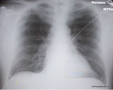
The symptoms have worsened over a 2-month period. Past medical history is unremarkable. Here, review the ED chest x-ray film and ECG. Do you see any clues to a diagnosis?
Figure 1. Plain film of chest. (click each image to enlarge)

Figure 2. ECG tracing in emergency department.

A 56-year-old man presents to the emergency department (ED) with gradually worsening dyspnea on exertion of 2 months’ duration. He has had an occasional dry cough but no fever, chest pain or discomfort, leg swelling, melena, or other symptoms. He states he didn’t come sooner because a few times he felt as though the symptoms were resolving and thought he would get over it. But each time the shortness of breath became worse again. He reports that today it is particularly bad and he couldn’t even walk up the 10 stairs to his apartment without getting severely short of breath. At rest, he says, the symptoms abate. He has never experienced anything like this.
Although he does smoke and drink socially, he denies any past medical history except for psoriasis and is not on any medications. He states his father died of a heart attack in his mid-60s and his mother died of a stroke at age 76. He has one sibling, a younger sister who has diabetes, but she has no heart disease.
On physical examination, he is afebrile with a pulse of 108 beats/min; blood pressure, 118/70 mm Hg; respiration, 22 breaths/min; and a pulse oximetry of 94% on room air. His head and neck exam is normal without jugular venous distention. His lungs are clear without rales or wheezing. His heart is regular without murmur and his legs have no swelling, tenderness, or cords. The rest of his physical exam is normal.
Blood work is ordered, including a CBC, metabolic panel, and cardiac troponin-I, which are all normal. His chest x-ray film (Figure 1) and ECG tracing (Figure 2) are shown at left above.
What is the most likely diagnosis?
What other conditions should be in your differential?
Is his presentation “typical” or “atypical”?
Are there any specific “pitfalls” demonstrated by this case?
Please leave your comments below and then click here for answers and discussion.
