- Clinical Technology
- Adult Immunization
- Hepatology
- Pediatric Immunization
- Screening
- Psychiatry
- Allergy
- Women's Health
- Cardiology
- Pediatrics
- Dermatology
- Endocrinology
- Pain Management
- Gastroenterology
- Infectious Disease
- Obesity Medicine
- Rheumatology
- Nephrology
- Neurology
- Pulmonology
Drug-Induced and Other Lung Diseases: A Photo Essay
Drug-induced hypersensitivity pneumonitis, drug-induced organizing pneumonia, radiation-induced lung injury, allergic fungal rhinosinusitis, pulmonary tuberculosis, empyema-this week’s slide show sheds light on several presentations.

Case 1:
More than 350 agents are associated with iatrogenic lung diseases. Drug-induced lung diseases (DILDs) vary in their pathophysiology, presentation, and prognosis. Proper diagnosis requires a high index of suspicion and familiarity with associated clinical syndromes. DILD typically is a diagnosis of exclusion of alternative causes of respiratory illness.
This lung biopsy specimen shows loosely formed granulomas near the terminal bronchioles and lymphocytic and plasma cell infiltration of the alveolar walls, indicating drug-induced hypersensitivity pneumonitis.
This clinically distinct pulmonary syndrome is characterized by a complex immunological reaction. Patients may have acute, subacute, or chronic fevers, chills, malaise, and dyspnea. Pulmonary function tests demonstrate a reduced FVC and carbon monoxide–diffusing capacity.
High-resolution CT (HRCT) of the chest may reveal bilateral upper lobe–predominant ground-glass opacities, poorly defined centrilobular nodules, and air trapping on expiratory scans. In chronic cases, radiographic and HRCT features may progress to fibrotic changes.
Drugs associated with a hypersensitivity pneumonitis include nitrofurantoin and methotrexate.
Case and image contributed by Krish Bhadra, MD, and Benjamin T. Suratt, MD.
NEXT CASE »
For the discussion, click here.
Case 2:
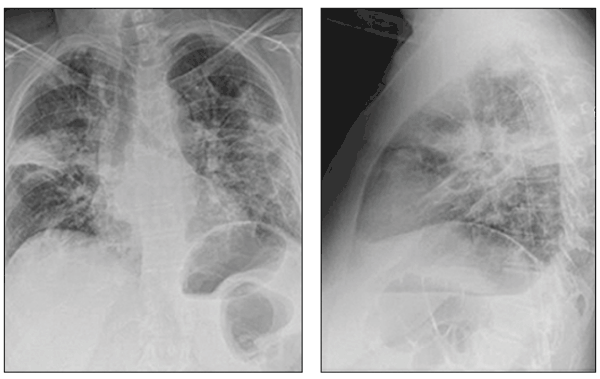
Multifocal airspace opacities can be seen on these chest radiographs from a patient with drug-induced organizing pneumonia, a unique form of lung disease.
Patients with organizing pneumonia may present with a flu-like illness, with fever, cough, weight loss, and progressive dyspnea. Bronchiolitis obliterans once had been considered a major feature.
Chest radiographs demonstrate patchy areas of consolidation, ground-glass and nodular opacities, and bronchial wall thickening and dilation.
For diagnosis, drug-induced organizing pneumonia often requires lung biopsy, which demonstrates preserved lung architecture with distinct intraluminal buds of granulation tissue.
Agents associated with drug-induced organizing pneumonia include amiodarone, bleomycin, and carbamazepine.
Case and images courtesy of Krish Bhadra, MD, and Benjamin T. Suratt, MD.
NEXT CASE »
For the discussion, click here.
Case 3:
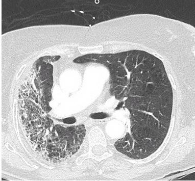
An 84-year-old woman presented to the ED with progressive dyspnea and hypoxemia. Her oxygen saturation was 84% on room air and the rest of her vital signs were within normal limits. She had had a dry, nonproductive, hacking cough for a few months without hemoptysis. She denied chest pain, syncope, fevers or chills.
The patient’s past medical history was significant for squamous cell skin cancer of the right shoulder. Treatment included surgical excision followed by 7 weeks of radiation therapy. Results of a CT scan of the chest are shown.
Radiation-induced lung injury after radiation therapy for squamous cell skin cancer was the diagnosis. Incidental radiation-induced lung injury often is dependent on dose of radiation and generally is unavoidable. Acute radiation pneumonitis may develop and can present with a dry cough and dyspnea. Pulmonary fibrosis also may cause dyspnea but is the late complication.
Radiographic imaging and pulmonary function testing can identify the degree of lung injury.
Image and case provided by Jonathan S. Ilowite, MD, and Roman Spivak, MD.
NEXT CASE »
For the discussion, click here.
Case 4:
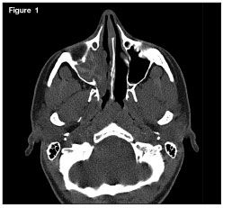
An 18-year-old woman with a history of allergic rhinitis and moderate persistent asthma presented with right-sided nasal congestion. Her symptoms persisted even with her usual allergy medications, allergen immunotherapy, and 2 courses of antibiotics. A CT scan showed complete opacification of the right maxillary sinus with increased attenuation of the mucin.
The patient had functional endoscopic sinus surgery, which was significant for thick mucin and nasal polyposis. Bacterial cultures of the mucin were negative; fungal cultures grew Curvularia.
Her diagnosis was allergic fungal rhinosinusitis, the most common form of noninvasive fungal sinusitis. This condition should be considered in any atopic patient who presents with chronic sinusitis refractory to usual antibiotic therapy.
The diagnosis is based on surgically obtained allergic mucin; allergic mucin demonstrating fungal hyphae or sinus culture, positive for a fungus; no evidence of invasive disease, necrosis, granulomas, or giant cells; and exclusion of other fungal sinus disorders.
Case and image contributed by Joseph J. Sclafani, MD, Douglas Gottschalk, MD, and Kirk H. Waibel, MD.
NEXT CASE »
For the discussion, click here.
Case 5:
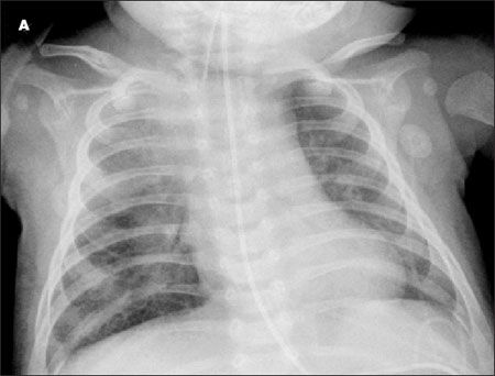
A 10-week-old baby girl with a history of difficulty in breathing presented with stridor, tachypnea, wheezing, and increased work of breathing. This case emphasizes the difficulty in making a diagnosis of pulmonary tuberculosis (TB) in an infant who has a nonspecific presentation and no travel or contact history.
The girl’s mother first noted abnormal breathing sounds at about age 4 to 6 weeks. At age 9 weeks, she was hospitalized for possible sepsis. A chest radiograph showed nonspecific increased bronchovascular markings. A viral syndrome was the diagnosis.
A chest radiograph showed a hyperinflated right lung with a shift of the mediastinal contents into the left hemithorax, suggestive of air trapping and consolidation in the right upper lobe and hilar region; there was no thymic shadow. A CT scan of the chest showed diffuse mediastinal and right hilar adenopathy; this caused partial compression of the right main-stem bronchus. MRI showed bilateral pleural effusion with hilar lymphadenopathy and mediastinitis. Examination of bronchial alveolar lavage fluid by PCR and culture was positive for Mycobacterium tuberculosis.
The diagnosis of infant TB is complicated by the lack of a clear response to a tuberculin skin test, absence of a gold standard diagnostic test, and difficulty in collecting respiratory specimens.
Case and image provided by Christopher Young, MD, Charelle Lockhart, MD, Luis Rodriguez, MD, and Akram Tamer, MD.
NEXT CASE »
For the discussion, click here.
Case 6:
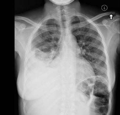
A 33-year-old woman with sickle cell disease was admitted with a 2-day history of high fevers, chills, and severe right-sided pleuritic chest pain. She had been well until the previous week, when upper respiratory–type symptoms developed. The symptoms progressed to severe right-sided pleuritic pain, a cough productive of green phlegm, a high temperature, and fatigue. A radiograph and a CT scan of the chest were obtained.
Because patients with sickle cell disease have functional asplenia, they are predisposed to severe infection, particularly with encapsulated organisms. Empyema would be a logical choice for this patient’s presentation.
Case and image courtesy of Jonathan Ilowite, MD.
