- Clinical Technology
- Adult Immunization
- Hepatology
- Pediatric Immunization
- Screening
- Psychiatry
- Allergy
- Women's Health
- Cardiology
- Pediatrics
- Dermatology
- Endocrinology
- Pain Management
- Gastroenterology
- Infectious Disease
- Obesity Medicine
- Rheumatology
- Nephrology
- Neurology
- Pulmonology
Differentiating Loxoscelism From Cutaneous Anthrax and Lyme Erythema Migrans
Loxoscelism is often misdiagnosed, in part because the clinical presentation of loxoscelism is similar to that of other conditions, such as cutaneous anthrax and Lyme erythema migrans (EM). Differentiating these disorders is important because some of these conditions require early treatment to achieve the best clinical outcomes. Unfortunately, using geography to make or exclude a diagnosis is becoming less reliable.
Loxoscelism is often misdiagnosed, in part because the clinical presentation of loxoscelism is similar to that of other conditions, such as cutaneous anthrax and Lyme erythema migrans (EM). Differentiating these disorders is important because some of these conditions require early treatment to achieve the best clinical outcomes. Unfortunately, using geography to make or exclude a diagnosis is becoming less reliable.
Loxoscelism
There are 6 species of brown spiders in the United States that can cause necrotic arachnidism.1,2 Of these, the most venomous is Loxosceles reclusus. As the name suggests, the brown recluse spider has a preference for caves, woodpiles, and other low-traffic, sheltered areas. Brown recluse spiders are commonly called fiddlebacks because of the upside-down violin-shaped marking under the mouthparts on their dorsum.
Historically, the brown recluse spider is indigenous to the upper Mississippi valley. However, this spider has reportedly been found in almost every continental state.3 These spiders can adapt to new environments, and it is not uncommon for a spider inhabiting a cardboard box or a stored sofa in a home in southern Missouri to survive a move to another state and infest an air-conditioned building.
The brown recluse spider is endemic to the area in which I have my practice (a rural area in southeastern Missouri). In 35 years, I have treated more than 100 patients with brown recluse spider bites. In my home, I have found brown recluse spiders even after routine pesticide spraying. Most of the brown recluse spider bites that I have treated have occurred in patients' homes rather than outdoors.
Brown recluse spiders are nonaggressive toward humans but will bite in self-defense if threatened. For example, bites can occur when a sleeping person rolls onto a spider that has found its way to the bed or when a person is putting on clothes that were left on the floor. The bites may be initially painless, but severe pain that is disproportionate to the appearance of the lesion can develop within 48 hours. Pain generally is the factor precipitating the office visit.
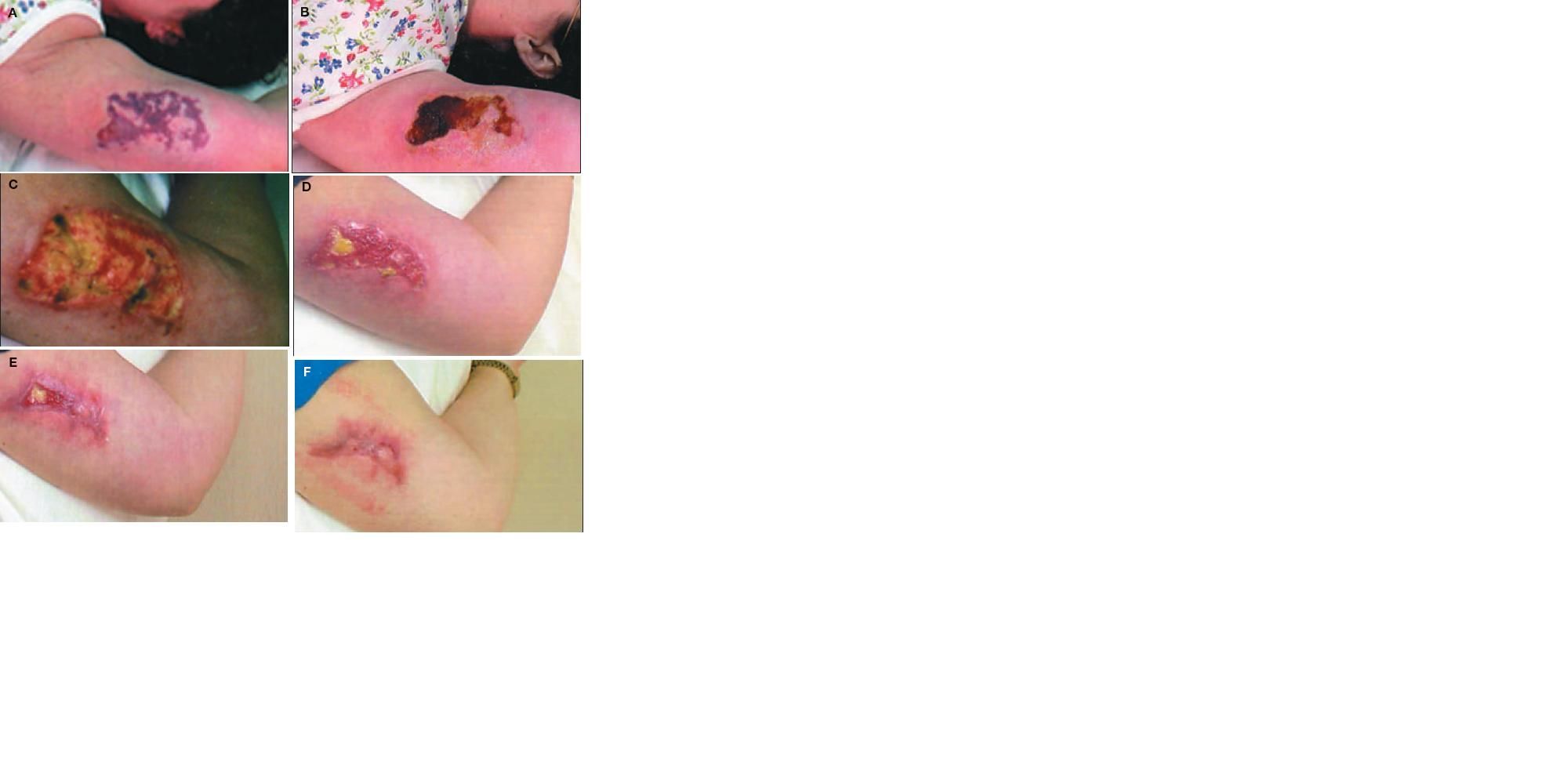
Brown recluse venom enzymes,such as sphingomyelinase D, cause red blood cells to leak into tissue spaces. The red blood cells, being relatively heavy, can produce a gravity-dependent erythema that can have a runny-egg appearance. The red, white, and blue sign, which is common in severe brown recluse envenomations, generally appears within 72 hours and is characterized by concentric zones of erythema outside of ischemia, which surrounds a smaller area of necrosis.4,5 The progression of severe loxoscelism is shown in Figure 1.

Figure 1
-
This patient presented with increasing pain in the left arm,which she noted on wakening. A spider bite was suspected, and she wasinstructed to return home and search her bedding for this spider. Thedead spider, apparently crushed by her arm, was recovered; it was identifiedas a brown recluse. On day 3 post-envenomation, increased erythemaand vesiculation and the red, white, and blue sign (erythema,ischemia, and necrosis) were evident (A). By day 8, an eschar hadformed (B). Photo C shows the lesion 1 week after debridement, on day23. By day 38, the patient reported negligible pain (D). Over the next5 weeks, the wound continued to heal (E, day 52), with less scarringthan expected (F, day 74).
Treatment is generally directed at pain relief, which can range from oral NSAIDs to hospitalization for administration of intravenous narcotics. I do not recommend intralesional corticosteroids, and surgical debridement is usually not warranted unless the eschar is severe.
Cutaneous anthrax
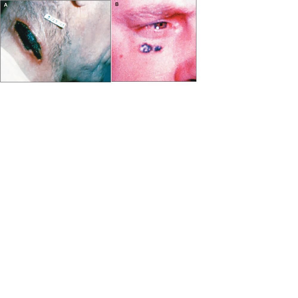
Historically, the primary risk factor for cutaneous anthrax is contact with animal parts or infected animals. If anthrax is used as a bioterrorism agent, however, anyone is at risk. Cutaneous anthrax is the most common type of anthrax infection. The lesions characteristically are painless and involve marked edema and the formation of an eschar (Figure 2). Other symptoms can include painful lymph nodes, fever, and headache. Treatment with penicillin, doxycycline, or ciprofloxacin is appropriate. The length of treatment is typically 60 days.

Figure 2
-
Lesions A and B were identified as cutaneous anthrax. Although quite rare in the United States, anthrax has garnered increased attention because of its otential use as a bioterrorism agent.
Lyme erythema migrans
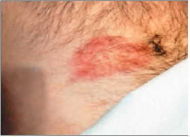
The most common manifestation of Lyme disease is EM. Lyme disease is caused by a bite from either an Ixodes scapularis or, on the West Coast, Ixodes pacificus infected with Borrelia burgdorferi. There is also a Lyme-like illness called southern tick-associated rash illness (STARI), or Masters disease.6 STARI can mimic Lyme disease-associated EM (Figure 3), and its cause is uncertain.7-10 Vectored by Amblyomma americanum (the Lone Star tick),11 STARI is primarily seen in the midwestern and southern states. The range of the Lone Star tick is spreading; it has been found in the Northeast12 all the way to Maine.13

Figure 3
-
This patient has southern tick-associated rash illness (STARI), also known as Masters disease. This Lyme disease mimic is vectored by
Amblyomma americanum.
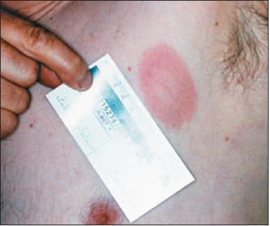
The classic appearance of Lyme EM is an expanding annular erythematous lesion that may have a classic bulls-eye appearance occurring 2 to 21 days after a bite from a tick infected with B burgdorferi (Figure 4). However, atypical presentations are common, and any lesion 5 cm or larger should be viewed with suspicion, especially if the patient has a history of frequent tick exposure without previously having had a similar-looking rash.

Figure 4
-
This patient, with culture-confirmed Lyme disease, has Lyme erythema migrans; the classic bulls-eye rash is evident.
Lyme EM is associated with minimal pain and edema. Although necrosis rarely occurs in Lyme EM, the rashes resulting from a tick bite and a brown recluse spider bite can appear similar, often confounding the diagnosis.
Both Lyme EM and Lyme-like lesions are typically treated with a 10- to 30-day course of oral antibiotics, usually doxycycline, 3 mg/kg in divided doses; cefuroxime, 500 mg bid; or amoxicillin, 500 mg tid. If there is evidence of early dissemination, such as fever, flu-like illness, severe headache, multiple lesions, or lymphadenopathy, the longer durations of treatment are recommended.
Discussion
Of these diagnoses, loxoscelism is the easiest to characterize clinically, with the punctum of a brown recluse spider bite usually being eccentric or off center. Edema can
