- Clinical Technology
- Adult Immunization
- Hepatology
- Pediatric Immunization
- Screening
- Psychiatry
- Allergy
- Women's Health
- Cardiology
- Pediatrics
- Dermatology
- Endocrinology
- Pain Management
- Gastroenterology
- Infectious Disease
- Obesity Medicine
- Rheumatology
- Nephrology
- Neurology
- Pulmonology
Differentiating Common Annular Lesions: Tinea Corporis vs Granuloma Annulare
Tinea corporis typically presents as an annular erythematous plaque with a raised leading edge and scale. Granuloma annulare classically presents with 1 or more indurated, erythematous or violaceous annular plaques on the extremities. Here, more on diagnosis and treatment.
A 51-year-old woman presented to her primary care physician with a self-diagnosis of “ringworm” on the dorsum of her left hand and her right posterior calf. The nonpruritic, violaceous, annular, and erythematous plaques were diagnosed as tinea corporis and clotrimizol antifungal cream was prescribed. The rash persisted despite 2 weeks of treatment and the patient was referred to a dermatologist.
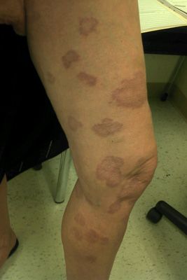
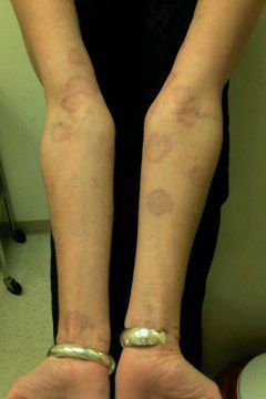
A biopsy from the lesion on the hand was consistent with features of localized granuloma annulare. The patient was reassured that the condition was self-limited. The lesion on her calf, however, grew larger and new nummular and annular plaques developed that ultimately involved her legs, arms, abdomen, and back.
A new diagnosis was made of generalized granuloma annulare. During the next 2½ years, the patient received numerous systemic and topical treatments with little improvement in symptoms. Treatments included topical corticosteroids, intralesional and intramuscular injections of triamcinolone, biotin (vitamin H, coenzyme R), hydroxychloroquine, quinacrine, pentoxifylline, and dapsone. She is currently receiving treatment with methotrexate and folic acid, although the response has been poor.
Tinea corporis and granuloma annulare are commonly seen in primary care practice. They have similar presentations, and subtle differences, and so are often misdiagnosed. A missed or erroneous diagnosis can lead to initial treatment failure and/or unnecessary therapy.
Tinea corporis-a dermatophyte infection- manifests on the trunk and extremities (excluding the hair, nails, palms, soles, and groin) and often mimics benign inflammatory skin conditions, such as granuloma annulare. Tinea is generally limited to the stratum corneum and is most commonly caused by Trichophyton rubrum and Trichophyton mentagrophytes.1 The infection can be transmitted from human to human, from animal to human, and from soil to human.2
Tinea corporis typically presents as an annular erythematous plaque with a raised leading edge and scale. Clearance occurs in the center of the lesion; however, residual nodules may be scattered throughout the infected area. This creates the characteristic annular plaque that gives the disease its common name, “ringworm.” Tinea lesions also can appear as arcuate, circinate, and oval.2
When you suspect tinea corporis infection, scrape the periphery of lesions to harvest surface scale for a KOH preparation. Fungal cultures on Sabouraud agar medium may also be used to confirm the diagnosis.3
Topical azole creams-clotrimizole or miconazole-are first-line therapy for tinea corporis. Second-line topical therapies include terbinafine 1% cream, ciclopirox, and 40% urea cream.4
Granuloma annulare is a benign inflammation that classically presents with 1 or more indurated, erythematous or violaceous annular plaques on the extremities. Unlike tinea corporis, scale is absent and the lesion may or may not be pruritic. Although its etiology is unknown, 4 clinical variants have been identified:
• Localized
• Disseminated
• Subcutaneous
• Perforating
While diagnosis can be made by visual examination, definitive diagnosis requires skin biopsy. Lesions are histologically characterized by necrobiotic collagen surrounded by a lymphohistiocytic infiltrate. Based on the T-cell subpopulations identified in the infiltrate, a delayed-type hypersensitivity reaction to an unknown antigen has been postulated as the precipitating event. Factors suspected in triggering granuloma annulare include trauma, insect bite reactions, tuberculin skin testing, sun exposure, PUVA therapy, and viral infections.1

Figure 1
The localized form of granuloma annulare usually resolves spontaneously, generally running a course of up to 2 years,5 with no long-term sequelae. Reassurance is usually all that is necessary. Treatment options include intralesional or topical corticosteroids and cryotherapy. However, granuloma annulare may progress from localized disease to the disseminated form, as was the case with the patient in this case history.
Disseminated granuloma annulare may be persistent, lasting for as few as 3 years but in some cases up to 10 years.5 Patients often seek active treatment for cosmetic reasons, often with disappointing results. Current treatment guidelines suggest a wide range of possibilities: PUVA with oral or topical psoralens, UVA alone, etretinate and isotretinoin, topical tacrolimus, dapsone, cyclosporine, systemic corticosteroids, chlorambucil, antimalarials, potassium iodide, pentoxifylline, nicotinamide (niacinamide), topical vitamin E, fumaric acid esters, defibrotide, doxycycline, hydroxyurea, and pulsed dye laser.3
Many reports supporting or refuting the association of granuloma annulare with diabetes mellitus have been published.1 In one retrospective study of 84 patients, 12% were found to have diabetes mellitus, and these patients were more likely to suffer from chronic relapsing granuloma annulare than were non-diabetic patients.6 Therefore, in patients presenting with granuloma annulare, screening for diabetes mellitus should be considered.
Granuloma annulare has also been associated with solid organ tumors, Hodgkin disease, and non-Hodgkin lymphoma and therefore has been considered to be a paraneoplastic reaction. However, in these patients, the clinical presentation is frequently atypical, with painful lesions in unusual locations, including the palms and soles.1

Figure 2
Other diagnostic considerations
Other, less common annular skin conditions that should be considered in a differential diagnosis include7:
• Pityriasis rosea, a self-limited papulosquamous skin eruption, is characterized by small, fawn-colored lesions distributed along skin cleavage lines. The “herald patch” may appear as an annular lesion with an erythematous raised border, fine scale, and central clearing.
• Urticaria may present at non-scaling annular plaques and is characterized by pruritic, well-circumscribed erythematous lesions that last between 90 minutes and 25 hours.
• Cutaneous sarcoidosis is an idiopathic, multisystem granulomatous disease that is characterized by non-caseating granuloma formation in virtually any organ system. Sarcoidosis may present as coalescing papules forming annular, indurated plaques.
• Hansen disease (leprosy) is transmitted by the acid-fast bacillus Mycobacterium leprosae. It may present as one or more annular, sometimes scaly, plaques that may lack sensation.
• Subacute cutaneous lupus erythematosus can present in annular or papulosquamous form. Photosensitivity is a hallmark, and lesions usually present on sun-exposed areas of the skin.
• Erythema annulare centrifugum typically presents as non-indurated annular patches with associated trailing scale inside erythematous borders. The lesions most commonly affect the trunk, buttocks, thighs, and legs.
• Erythema chronicum migrans is the cutaneous hallmark of Lyme disease. It is characterized by large erythematous patches that may appear anywhere on the surface of the skin. The lesions expand centrifugally and have central clearing leading to annular erythematous patches.
• Nummular eczema is associated with xerosis and is characterized by coin-shaped erythematous papules or plaques most commonly on the legs. Sometimes the lesions expand with central clearing, giving rise to annular plaques.
• Psoriasis is most commonly found on extensor surfaces and is characterized by erythematous thick plaques with silvery scale. However, psoriasis can arise as annular lesions with silvery scale only on the borders.
Teaching Points:
• Tinea corporis and granuloma annulare have similar presentations and subtle differences, and so are often misdiagnosed.
• Tinea corporis typically presents as an annular erythematous plaque with a raised leading edge and scale. Diagnosis is confirmed with KOH preparation or fungal culture.
• Granuloma annulare classically presents with 1 or more indurated, erythematous or violaceous annular plaques on the extremities. Diagnosis is confirmed by skin biopsy.
References:
References:
1. Bolognia JL, Jorizzo JL, Rapini RP, eds. Dermatology. 2nd ed. Spain: Mosby Elsevier; 2008:278, 1139-1140, 1426-1429.
2. Gupta AK, Chaudhry M, Elewski B. Tinea corporis, tinea cruris, tinea nigra, and piedra. Dermatol Clin. 2003;21:395–400.
3. Lebwohl MG, Heymann WR, Berth-Jones J, Coulson I, eds. Treatment of Skin Disease: Comprehensive Therapeutic Strategies. 3rd ed. China: Elsevier Limited; 2010:275-278, 736-745.
4. Weinstein A, Berman B. Topical treatment of common superficial tinea infections. Am Fam Physician. 2002;65:2095-2102. http://www.aafp.org/afp/2002/0515/p2095.html
5. Cyr PR. Diagnosis and management of granuloma annulare. Am Fam Physician. 2006;74:1729-1734. http://www.aafp.org/afp/2006/1115/p1729.html
6. Studer EM, Calza AM, Saurat JH. Precipitating factors and associated diseases in 84 patients with granuloma annulare: a retrospective study. Dermatology. 1996;193:364-368.
7. Hsu S, Le EH, Khoshevis MR. Differential diagnosis of annular lesions. Am Fam Physician. 2001;64:289-296. http://www.aafp.org/afp/2001/0715/p289.html
