- Clinical Technology
- Adult Immunization
- Hepatology
- Pediatric Immunization
- Screening
- Psychiatry
- Allergy
- Women's Health
- Cardiology
- Pediatrics
- Dermatology
- Endocrinology
- Pain Management
- Gastroenterology
- Infectious Disease
- Obesity Medicine
- Rheumatology
- Nephrology
- Neurology
- Pulmonology
Diastematomyelia
A 42-year-old woman had a "life-long" history of mild to moderate low back pain, without radiation, anesthesia, weakness, or incontinence. She denied recent trauma or an inciting event. The pain was constant and worse at the end of the day and with prolonged activity. Physical therapy, NSAIDs, tramadol, opioids, and manipulation provided minimal relief.

A 42-year-old woman had a "life-long" history of mild to moderate low back pain, without radiation, anesthesia, weakness, or incontinence. She denied recent trauma or an inciting event. The pain was constant and worse at the end of the day and with prolonged activity. Physical therapy, NSAIDs, tramadol, opioids, and manipulation provided minimal relief.
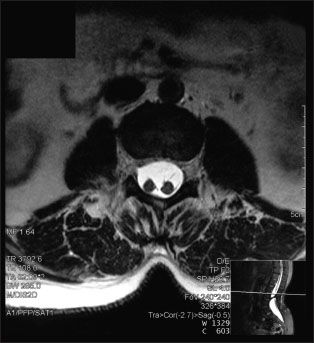
An MRI scan without contrast of the lumbar spine revealed a sagittal division of the spinal cord into 2 halves at the L2-3 level separated by a cartilaginous septum.
Diastematomyelia is a rare congenital neural-tube defect, in which a bony spicule or fibrous band from the body of one of the thoracic or lumbar vertebrae protrudes into the spinal canal, dividing the cord into 2 halves of varying lengths. In contrast, diplomyelia, in which the spinal cord does not reunite beyond the spur, results in complete division of the spinal cord with each half in possession of its own dural sac and nerve roots.1 Split spinal cords, of any degree, are usually accompanied by other skeletal anomalies, such as spina bifida, kyphoscoliosis, and hemivertebra, as well as varying degrees of spinal cord dysfunction. As the patient grows, a traction myelopathy may develop, and pain and progressive sensory, motor, and bladder symptoms may present in adulthood.2 However, most cases are diagnosed in childhood.
In most adult cases, symptoms are precipitated by specific circumstances, such as a traumatic event. Pain, often involving the analperineal region, is the most common complaint. Bilateral leg weakness and urological complaints are also common. Consider diastematomyelia in the presence of scoliosis; skin changes at the base of the spine; progressive foot deformities; calf and foot atrophy; and bowel, bladder, and gait disturbances.3 In patients with these abnormalities, surgery may be performed to untether the spine and prevent worsening neurological symptoms.
Surgery is indicated in adults with diastematomyelia when stenosis or cord tethering causes neurological deficits. With the exception of scoliosis, prophylactic spur removal is not recommended in asymptomatic adults.2,3
The patient was referred to neurosurgery and is awaiting consultation.
References:
REFERENCES:
1.
Ropper AH, Brown RH. Developmental diseases of the nervous system. In: Ropper AH, Brown RH, eds.
Adams and Victor's Principles of Neurology
. 8th ed. New York: McGraw-Hill Medical Publishing; 2005:chap 38.
2.
McDermott MW, Kunwar S, Berger MS. Neurosurgery and surgery of the pituitary. In: Doherty GM, Way LW, eds.
Current Surgical Diagnosis & Treatment
. 12th ed. New York: Lange Medical Books/McGraw-Hill; 2006:chap 37.
3.
Lewandrowski KU, Rachlin JR, Glazer PA. Diastematomyelia presenting as progressive weakness in an adult after spinal fusion for adolescent idiopathic scoliosis.
Spine J
. 2004;4:116-119.
