- Clinical Technology
- Adult Immunization
- Hepatology
- Pediatric Immunization
- Screening
- Psychiatry
- Allergy
- Women's Health
- Cardiology
- Pediatrics
- Dermatology
- Endocrinology
- Pain Management
- Gastroenterology
- Infectious Disease
- Obesity Medicine
- Rheumatology
- Nephrology
- Neurology
- Pulmonology
Cutaneous Leukocytoclastic Vasculitis
This hypersensitivity reaction may be secondary to medications, infection, collagen-vascular disorders, or an occult malignancy. When it is localized to the skin, prognosis is excellent.
Figure 1. (Click on all figures to enlarge)
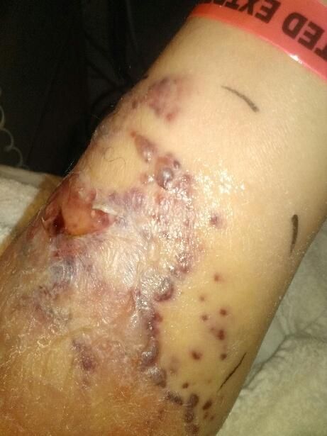
Figure 2.
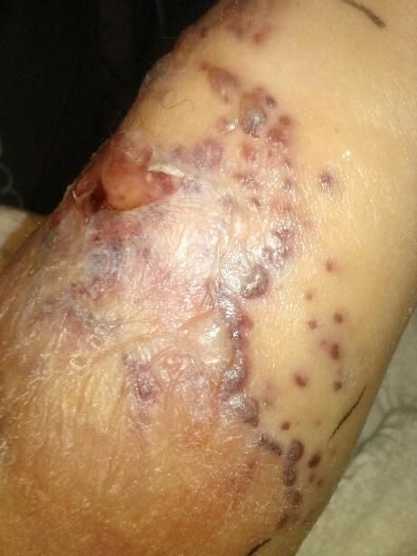
Figure 3.
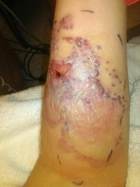
Figure 4.
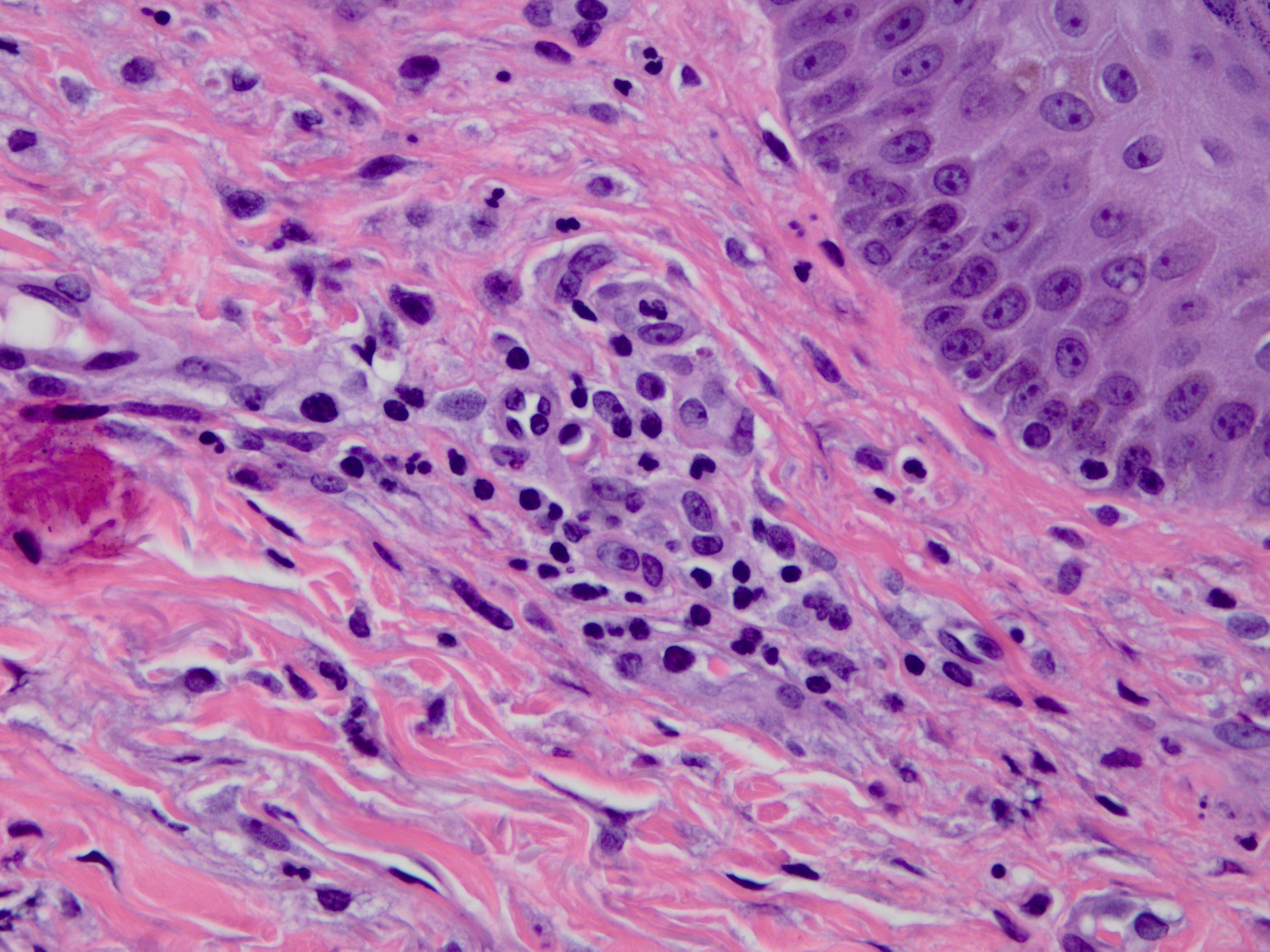
Figure 5.
A 55-year-old man with a past medical history of chronic lymphocytic leukemia (CLL) presented to the hospital with a four-month history of fever, chills, night sweats, and fatigue. During his admission, a bone marrow biopsy revealed that monoclonal lymphocytes accounted for approximately 75% of bone marrow cellularity. He received his first chemotherapy cycle of bendamustine, rituximab, and pegfilgrastim as an inpatient and was discharged home three days later. He followed up with his oncologist as an outpatient and during the next week received pegfilgrastim for chemotherapy-induced neutropenia and IV fluids for dehydration. He was also started on prophylactic acyclovir and a short course of allopurinol.
Two days after receiving IV fluids he reported significant pain, erythema (approximately 12 cm in length), bullous lesions, induration, and edema at the IV infusion site on his left forearm (Figures 1, 2, and 3) with erythematous lymphatic tracking proximally towards his axilla. He was referred back to the hospital and admitted for suspected cellulitis. On admission he was found to be afebrile with a WBC count of 2600/µL and ANC of 1.9 x10⁹/L. Treatment was initiated with IV vancomycin and IV piperacillin-tazobactam and blood cultures were drawn. There was concern for a possible deep vein thrombosis (DVT). Venous ultrasound, however, was negative for an abscess or venous thrombosis. CT scan and MRI results also were consistent with cellulitis with no evidence of an abscess or osteomyelitis.
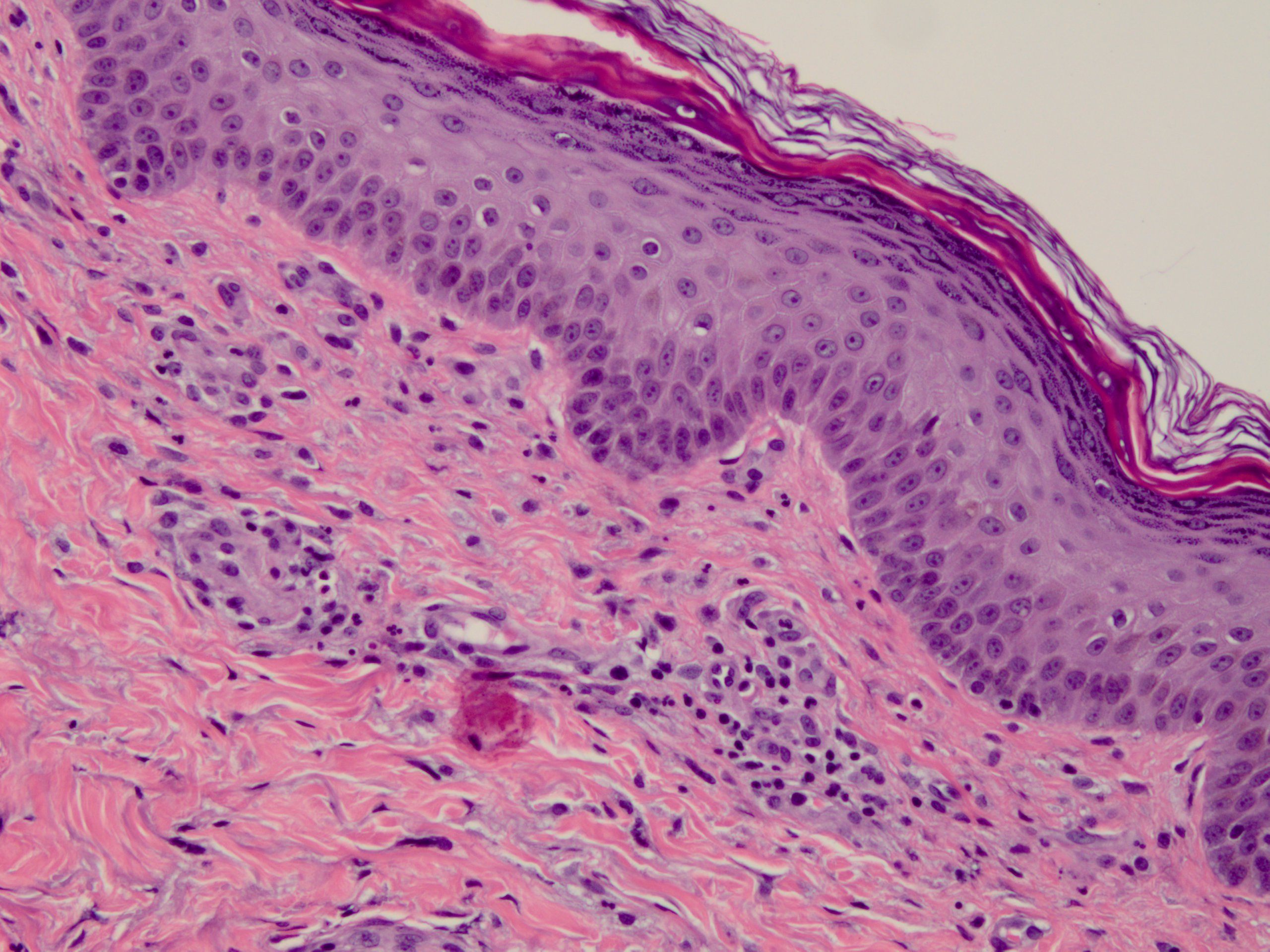
The patient’s pain, erythema, and edema continued to worsen despite 72 hours of treatment with broad spectrum IV antibiotics. The possibility of Sweet syndrome or pemphigoid was considered so a punch biopsy of the bullae was obtained and sent to pathology for analysis. The preliminary pathology report revealed a mixed, predominantly neutrophilic, perivascular infiltrate obliterating vessel walls, leukocytoclasis, erythrocyte extravasation, and overlying epidermal necrosis. The histologic findings supported a diagnosis of late stage cutaneous leukocytoclastic vasculitis (LCV). Antibiotics were discontinued and the patient was immediately started on IV methylprednisolone sodium succinate. Within 24 hours a significant decrease in erythema and edema was clearly visible and the patients’ pain was markedly improved. IV steroids were continued for another 48 hours. The patient continued to improve and was eventually discharged on a 2-week tapered course of oral prednisone.
Discussion
Cutaneous LCV, also known as hypersensitivity vasculitis, is a histopathologic term used to describe a small-vessel vasculitis. Leukocytoclasis refers to vascular damage caused by nuclear debris from infiltrating neutrophils1 (Figures 4 and 5). The condition may be secondary to medications, bacterial infection, collagen-vascular disorders, or an underlying malignancy. Approximately half of all cases, however, are idiopathic with no identifiable etiology.2 The incidence of LCV is presumed to be quite low. A recent study published by the Mayo Clinic found the incidence to be 4.5 per 100,000 persons per year.3 LCV occurs more often in Caucasians than in any other race and affects men and women equally. It is mostly an acute phase reaction but approximately 10% of patients will have chronic or recurrent disease. LCV may be localized to the skin or may be associated with systemic involvement. In the absence of internal organ involvement, the prognosis is excellent, with most cases resolving within several weeks.
Diagnosis. “When there is an issue… get tissue.” A punch biopsy of a new lesion should be performed in most adult patients suspected of having LCV.
Treatment. If exposure to a new drug is the likely etiology then a biopsy may not be necessary, as long as the lesions resolve within a few weeks. Patients with an identifiable cause should receive treatment for that cause. Stopping a drug suspected of causing LCV can result in rapid clearing of the inflammatory process.4 Common first-line treatments for cutaneous LCV are colchicine and dapsone. A combination of these agents can also be utilized when one or the other alone is insufficient. Corticosteroids are strongly recommended, especially with bullous type lesions, in order to prevent cutaneous ulcerations and scarring. Patients with severe or debilitating LCV may require high doses of corticosteroids. A course of an immunosuppressive agent also should be considered for refractory or severe disease (eg, cyclophosphamide, azathioprine, methotrexate, mycophenolate, mofetil, or rituximab).5
Take-home Points
Cutaneous LCV should be considered as a differential diagnosis when cutaneous manifestations present after a new drug is administered or in the case of an underlying malignancy. Once again, when there is an issue, get tissue. A tissue biopsy can confirm the diagnosis.
