- Clinical Technology
- Adult Immunization
- Hepatology
- Pediatric Immunization
- Screening
- Psychiatry
- Allergy
- Women's Health
- Cardiology
- Pediatrics
- Dermatology
- Endocrinology
- Pain Management
- Gastroenterology
- Infectious Disease
- Obesity Medicine
- Rheumatology
- Nephrology
- Neurology
- Pulmonology
Cryptococcal Meningitis: Review of Current Disease Management
The incidence of cryptococcal infections in the HIV-infectedpopulation has diminished because of the effectiveness of anti retroviraltherapy, whereas the incidence in non–HIV-infectedhosts has grown. Despite improvements in antifungal therapy,successful outcomes in the management of cryptococcalmeningitis are dependent on a high index of clinical suspicion,appropriate use of diagnostic assays, early and aggressiveantifungal therapy, and recognition of complications such asincreased intracranial pressure and immune reconstitutionsyndromes. Published guidelines for the care of patients withcryptococcal meningitis are available and may be adapted toindividual patient requirements. Basic and clinical studies areneeded to further define the components of immune protection,optimal therapy in special patient populations, and the recognitionand treatment of complications of cryptococcal meningitis.[Infect Med. 2008;25:11-23]
Despite improvements in overall disease-free survival, invasive fungal infections continue to play a prominent role in health issues faced by immunocompromised patients. Effective HAART in patients with HIV infection has decreased the incidence of fungal infections, including those caused by Cryptococcus neoformans. Cryptococcal meningitis remains an important infectious syndrome among the growing population of chronically immunodeficient patients who undergo solid organ or hematopoietic stem cell transplantation and presents a unique diagnostic and therapeutic challenge for clinicians.1
MICROBIOLOGY
There are close to 20 known species of Cryptococcus. C neoformans is the most common human pathogen. It exists as an encapsulated yeast in the environment, occurring in 4 different serotypes determined by capsular agglutination reactions and designated as A, B, C, and D. Serotypes A and D have historically been designated as "variety neoformans" (C neoformans var neoformans), although serotype Ais often placed in its own separate variety (ie, C neoformans var grubii).2
Serotypes B and C have been identified as C neoformans var gattii. Some authors, however, have proposed that they be placed into a separate species altogether.3 Historically, Cryptococcus gattii was associated with tropical and subtropical climates, but outbreaks of infection in humans and animals have been identified in the United States and Canada.4 Isolation of this particular organism from different native tree species in the Pacific Northwest has suggested a potential common environmental source.5 C neoformans, on the other hand, shows no geographic predilection.
Microscopically, C neoformans is a spherical, encapsulated yeast both in tissue and on culture media. Budding is seen in vitro, although true hyphal forms are rarely appreciated. In tissue histology, the yeast cells often appear crescent- or cup-shaped and are readily visualized with calcofluor white, Giemsa, Gomori methenamine-silver, or even routine Gram stains (Figure 1). The capsule readily takes up mucicarmine and appears reddish pink. C neoformans grows on most culture media within 48 to 72 hours, forming white, mucoid colonies, depending on how much capsular polysaccharide production is maintained.6
Figure 1 -
High-power microscopic Gomori methenamine-silver stain of a resected pulmonarynodule from a kidney transplant recipient showing multiple cryptococcal yeast forms.
The thick capsule of Cryptococcus is thought to be an important virulence determinant. Although the yeast measures 4 to 6 ?m in diameter, capsule thickness varies from 1 to more than 30 ?m.7 Organisms are smaller and less well-encapsulated in the environment, allowing them to reach terminal airways following inhalation.
As the fungus enters the host via the respiratory tract, individual yeast cells can inhibit destruction by phagocytes and replicate efficiently. Cryptococcus strains that do not produce capsules-or that are hypocapsular- are more readily destroyed by phagocytic cells and are less virulent in animal models.8
Phenol oxidase is another important virulence determinant. This enzyme allows the yeast to produce melanin from phenolic compounds such as catecholamines. This may account for some aspects of the organism's neurotropism. Dopamine in the CNS provides Cryptococcus with a supply of melanin, which may protect the yeast from host defenses. Likewise, the potential toxic effects of accumulated catecholamines may be mediated by their conversion to melanin.9
EPIDEMIOLOGY
C neoformans is a fungus with a worldwide distribution that is found in the soil and commonly associated with bird droppings. The nitrogenous compounds in bird excrement serve as a source of nutrition and growth for the organism. Pigeons are most often implicated, although direct pigeon-to-human transmission has never been documented. One case involving a pet cockatoo and a kidney transplant recipient has been described, suggesting the possibility of animal-to-human transmission.10
Symptomatic infection in otherwise healthy persons is rare, particularly cases involving dissemination of the yeast beyond the lungs. Subclinical or only mildly symptomatic infection appears to be more common. In one study, conducted in New York, 70% of children aged 5 years and older had evidence of serological responses to Cryptococcus antigens, suggesting widespread exposure. 11 Inhalation of a large inoculum is thought to increase the risk of symptomatic infection in the immunocompetent patient. The virulence of certain strains of C neoformans also may play a role in determining whether infection develops in the setting of normal immunity.
Infection with C gattii has been associated primarily with immunocompetent patients, although underlying lung conditions and corticosteroid use are still thought to be risk factors.5 During an outbreak of C gattii infection on Vancouver Island, British Columbia, from 1999 through 2001, pneumonia was common, with meningitis occurring in approximately 20% of cases.5 Infection with this species has continued in the Pacific Northwest with a disease incidence as high as 36 cases per million people per year before 2006.5
By far, the majority of patients with cryptococcosis have a defect in cellular immune function. The most common underlying condition is AIDS. Patients who are receiving long-term corticosteroid therapy, have undergone solid organ transplantation (SOT), have an underlying malignancy, or have other defects in cellular immunity are also at risk. Previous reports indicated that cryptococcal infection occurs in more than 10% of patients with AIDS, although with the widespread use of effective HAART, the overall incidence appears to be declining.12 Despite this, the case-fatality rate appears stable at roughly 12% in HIVinfected patients.1 In areas of the world where HAART is less readily available and the incidence of AIDS is much higher, cryptococcal infection is more common.13
The exact incidence of infection caused by Cryptococcus in solid organ transplant recipients is not known. Previous surveys have suggested an overall incidence of slightly less than 3%.14 Mortality rates, at least in the short term, have been calculated at 14% overall in the SOT population.15
Liver transplant recipients, particularly those with preexisting immune defects, have an increased risk of complications from infection with C neoformans, and mortality approaches 40%.7 Liver transplant recipients at greatest risk for infection include those with abnormal cognition at presentation, underlying kidney failure, fungemia, or evidence of dissemination beyond the lungs. Use of calcineurin inhibitors as part of the post-transplantation immunosuppressive regimen may be partially protective, although this observation needs to be validated in prospective trials.15
Both tacrolimus and cyclosporine have the ability to inhibit C neoformans at higher temperatures in vitro and may affect the temperature sensitivity of the organism.16 Synergy between fluconazole and calcineurin inhibitors against Cryptococcus in vitro also has been noted,17 suggesting that the observed clinical effect may be multifactorial.
CLINICAL PRESENTATION
Infection with Cryptococcus starts with inhalation of the fungus. Pulmonary findings are an important and often underappreciated aspect of a disease dominated symptomatically by CNS involvement. Given widespread exposure, cryptococcal infections in immunosuppressed patients may be caused by reactivation of latent disease from the respiratory tract. Cough and chest pain are the most common presenting concerns in immunocompetent persons with symptomatic pulmonary cryptococcal infections. Other symptoms may include low-grade fever, night sweats, weight loss, and hemoptysis. 18 Some patients with isolated pulmonary disease are asymptomatic. 19 Concurrent disseminated disease occurs in about 15% of cases.20
Most immunocompromised patients with symptomatic cryptococcal infection present with fever and cough. In HIV-positive patients, the presentation of pulmonary cryptococcosis tends to be acute with severe symptoms such as fever, cough, dyspnea, and hypoxia.21 Retrospective data suggest the severity of the presentation is proportional to the deficit in CD4+ T cells and is greatest in those with CD4+ cell counts of less than 100/?L.22 Chest radiographic findings are variable but tend to show interstitial infiltrates.
In the non-HIV-infected immunosuppressed patient, pulmonary infection typically presents with well-defined nodules, often in the peripheral lung fields (Figure 2). Diffusely nodular or patchy alveolar infiltrates also may be seen, although cavitation, mediastinal adenopathy, pleural effusions, and calcification are rare.
Figure 2
-
Chest CT scan from a 41-year-old man with a history of diabetes mellitus type 1and kidney-pancreas transplant in whom an asymptomatic cryptococcal pulmonary nodule wasfound during workup for an unrelated complaint.
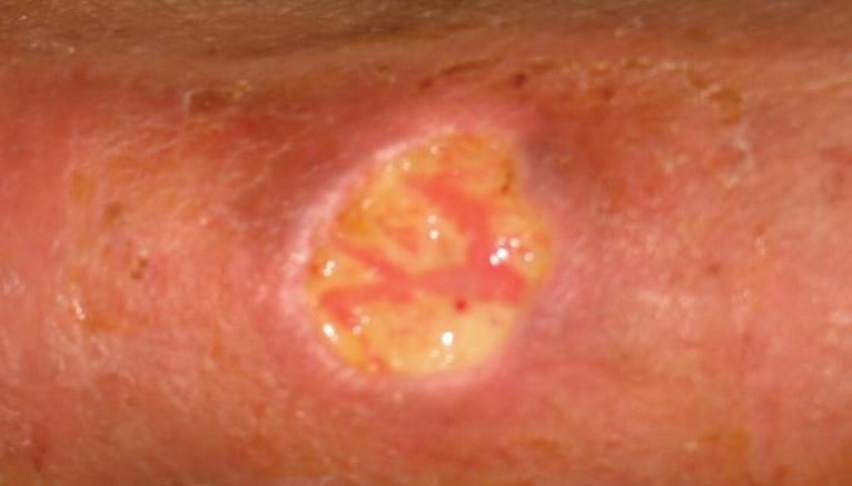
The most important clinical syndrome associated with cryptococcal infection in the immunosuppressed patient is meningitis. Once inhaled into the respiratory tract, the organism spreads via the bloodstream to other sites. Cases have been reported involving the skin (Figure 3), joints, bone, urinary tract and prostate, eye, cardiac tissue and pericardium, GI tract, peritoneum, thyroid, and larynx. The biological basis of the propensity of the organism for the CNS is unclear.

Figure 3
-
Abiopsy specimen from a kidney transplant recipient with persistent infection at thesite of an intravenous catheter revealed Cryptococcus neoformans. Subsequently, the patientwas found to have simultaneous pulmonary infection and asymptomatic meningitis.
In the immunocompromised patient without AIDS, infection may evolve over weeks to months before the diagnosis of meningitis is determined. The clinical syndrome in these patients tends to be similar to that of patients with AIDS and is marked by fever, headache, lethargy, and mental status changes. Symptoms may wax and wane. Early diagnosis requires a high index of suspicion. Focal neurological changes are uncommon, and patients may not experience the meningism, photophobia, or vomiting associated with other forms of meningitis. Increased intracranial pressure, however, with subsequent hydrocephalus and obstructive symptoms, is common in cryptococcal meningitis and in the immune reconstitution syndrome that may occur with reduction in immunosuppression. Patients may require repeated lumbar punctures to reduce intracranial pressure and may benefit from ventriculostomy or shunting procedures if persistent or severe hydrocephalus is present.23-25
Up to 30% of patients with AIDS and cryptococcal infection may exhibit an immune reconstitution inflammatory syndrome following initiation of HAART.26-28 This syndrome also has been reported in solid organ transplant recipients with tapering of immunosuppressive agents during therapy for acute infection. Clinical findings include relapse or worsening of meningitis with intracranial hypertension, hypercalcemia, cavitation of pulmonarylesions, intrathoracic lymphadenopathy, and abscess formation.29 These findings suggest that caution is required, particularly regarding corticosteroids, in the reduction of immunosuppression during the management of acute infection.
DIAGNOSIS
Isolation of the organism in culture is needed to definitively diagnose cryptococcal infection. In patients with cutaneous or metastatic disease, the organism may be isolated from cultures of respiratory tract specimens, blood, cerebrospinal fluid (CSF), and tissue biopsy samples. Isolation from sputum may occur in the absence of invasive disease and may be misleading. Conversely, bronchoscopic cultures and needle aspirates of focal lesions have a high yield even if fungal smear results are negative. We generally recommend lumbar puncture and CSF examination with pulmonary cryptococcal disease even in the absence of meningeal symptoms, in accordance with published guidelines.23 However, it should be noted that clinical trial data supporting such an approach are not available. In the case of meningitis, analysis of CSF may reveal only slight abnormalities in protein and glucose levels or cell counts. The latter tend to be more elevated in non-HIV-infected patients. Apredominance of mononuclear cells in the leukocyte differential of CSF is frequently seen. The organism is often easily cultured from the CSF, although both newer antigen assays and older microbiological techniques may provide a basis for early therapy.
India ink preparations of CSF samples may yield visible, distinctive organisms by clearly outlining the yeast and its large capsule. Although occasionally confused with artifact, organisms often can be identified by practiced examiners in between 25% and 50% of cases.30,31 Gram stains may be unable to distinguish yeast cells from host tissue. More recently, clinicians have relied on rapid protein assays that identify the Cryptococcus polysaccharide antigen. The Cryptococcus antigen assay can be performed on both CSF and serum samples and is useful for the diagnosis and management of cryptococcosis. Results are generally available before cultures give positive results.
Latex agglutination assays for Cryptococcus antigens in CSF samples have a sensitivity and specificity in the mid-90% range during meningitis.32 False-positive results have been reported with infections caused by Trichosporon beigelii and Capnocytophaga canimorsus, although the titer tends to be low in these infections (1:8 or lower).33,34 For the diagnosis of isolated pulmonary disease, a negative result from the serum assay can be misleading, and biopsy or culture may be necessary for accurate diagnosis.19,35 The sensitivity of serum Cryptococcus antigen testing in the setting of isolated pulmonary disease is unclear and may vary with the patient population, the clinical sample obtained, and the clinical laboratory performing the as say.
New serum assays for fungal infections such as the galactomannan assay, which was recently approved by the FDA for the early diagnosis of invasive aspergillosis, and the plasma (1->3)- ?-D-glucan assay are less well-studied in patients with cryptococcosis. Although an epitope of the C neoformans galactoxylomannan may be able to cross-react with As pergillus galactomannan assays,36 the test is not useful for the clinical diagnosis of cryptococcal infections. Clinical data suggest a sensitivity rate of the galactomannan assay for C neoformans to be below 15%37; because C neoformans does not release soluble (1->3)- ?-D-glucan in amounts high enough for routine detection,38 this assay is also not considered useful for clinical diagnosis. Thus, the latex agglutination assay for Cryptococcus specific antigens remains the test of choice despite some limitations.
THERAPY
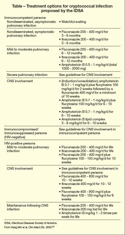
General guidelines for the treatment of cryptococcal infection, developed by the Infectious Diseases Society of America (IDSA) and published in 2000, are shown in the Table.23 (The document can be accessed at http://qcom.etsu.edu/intmed/divisions/ id/guidelines/Cryptococcal% 20infections.pdf).

Table
Immunocompetent persons with asymptomatic pulmonary disease without dissemination may not need therapy. These patients may still merit evaluation for occult immuno deficiencies. If treatment is desired in symptomatic non-immunosuppressed persons, fluconazole 200 to 400 mg/d for 3 to 6 months is recommended. Itraconazole 200 to 400 mg/d is an alternative. For patients with more severe infection, such as meningitis (ie, CNS involvement), amphotericin B and flucytosine are given. Treatment guidelines for meningitis should be followed in immunosuppressed patients.
Patients with meningitis should be treated regardless of the underlying integrity of the immune system. Any immunocompromised patient with evidence of cryptococcal infection should be treated. Therapy can be divided into 3 phases: induction, consolidation, and maintenance, although the specific components of therapy remain controversial.
In the immunosuppressed solid organ or hematopoietic transplant recipient, nephrotoxic agents, including amphotericin B and flucytosine, may be poorly tolerated; data on the ideal approach are lacking. The suggested dosage of flucytosine is 100 mg/kg/d, but in practice, many patients will not tolerate this dosage because of myelosuppression or other adverse effects. Patients with renal insufficiency are particularly susceptible to complications of therapy. Blood levels of flucytosine should be monitored.
When infection is limited to the lung, and no evidence of fungemia, dissemination, or CNS involvement is observed, induction monotherapy in the form of high-dose fluconazole (minimum 800 mg/d) may be an acceptable alternative in immunosuppressed solid organ transplant recipients. 39 Prospective data are lacking, however, and IDSA guidelines propose treating these patients like HIVinfected patients.23
The consolidation phase of therapy begins once the patient has shown clinical improvement after induction and can be switched to highdose fluconazole alone (defined usually as 400 mg/d for patients with normal renal and hepatic function). Fluconazole has been shown to be superior to itraconazole in preventing relapses in most patients.40 In vitro studies with newer azole antifungal agents such as voriconazoleand posaconazole have shown activity against isolates of C neoformans41,42 but, as yet, clinical data are lacking. Consolidation is often extended for at least 8 weeks if the patient does well clinically before reduction of the dose (to 200 mg/d) can be considered for maintenance and suppression.
Duration of maintenance therapy in non-HIV-infected immunosuppressed patients remains unclear but should be matched with the intensity of immunosuppression. IDSA guidelines recommend that maintenance therapy be continued for 6 to 12 months.23 According to prospective multicenter data on cryptococcal infection in this population, patients who received 6 months of maintenance therapy are at low risk for relapse overall,39 although clearly defined parameters remain elusive.
Induction therapy in HIV-positive patients generally starts with highdose amphotericin B (0.7 to 0.8 mg/kg/d) or liposomal amphotericin B (3 to 5 mg/kg/d) combined with flucytosine for 2 weeks, regardless of the site of infection. In meningitis, aggressive management of increased intracranial pressure is a key component of patient care. Increased intracranial pressures even in the setting of appropriate antifungal therapy can cause long-term neurological damage.43 The combination of amphotericin B with flucytosine is more rapidly fungicidal than amphotericin B combined with fluconazole or amphotericin B alone in cases of meningitis.44 Combining amphotericin B with flucytosine during this initial 2-week induction phase also is associated with significantly lower relapse rates in HIV-infected patients than when monotherapy is used.40
The duration of maintenance therapy remains a topic of debate. Historically, in the HIV-infected population, fluconazole maintenance therapy was continued indefinitely because of concerns about relapse in the setting of continued immunosuppression. Some data and clinical experience suggest that it is reasonable to consider discontinuation of maintenance therapy after patients have a response to HAART and have maintained CD4+ cell counts above 100 to 200/?L for an extended time.45 Occasional relapses have been noted in patients who still have CD4+ cell counts of less than 200/?L.46
Other antifungal agents such as the new echinocandins (caspofungin, micafungin, and anidulafungin) have no activity against Cryptococcus and, thus, have no role in therapy. Follow-up trials and reports using lipid formulations of amphotericin B have suggested efficacy and lower toxicity, making these preparations a viable alternative to standard formulations of amphotericin B.47-49 Adjunctive treatment in the form of interferon- ? with antifungal agents has been reported, although concerns about stimulating rejection in solid organ transplant recipients should be considered, making this therapy a last resort.50 After initiation of antifungal therapy, symptomatic patients are often slow to improve. As was described above, clinical symptoms may worsen after initial improvement both in patients with AIDS who are receiving HAART and in immunosuppressed patients who have reduction in the intensity of immunosuppression. 26,27 Immune reconstitution syndrome is thought to be caused by stimulation of a cytotoxic helper T-cell (TH1) response to infection.28 This kind of response in kidney transplant recipients also has been associated with a subsequent risk of rejection and graft loss and can be mistaken for a flare of the underlying infection.51
CONCLUSION
Although effective HAART has reduced the incidence of many opportunistic infections, such as cryptococcosis, in the HIV-infected population, increases in the incidence of infections in other immunocompromised hosts have highlighted the absence of a consensus regarding management of cryptococcosis in non-AIDS-affected immunocompromised hosts. A high index of suspicion must be maintained for this fungal infection. Untreated or insufficiently managed patients may have poor outcomes. Furthermore, studies are needed to investigate optimal therapies for acute infection in the non-AIDSaffected immunocompromised host, the use of adjunctive therapies, and the management of immune reconstitution syndromes and increased intracranial pressure in cryptococcal meningitis.
References:
- Warnock DW. Trends in the epidemiology of invasive fungal infections. Nippon Ishinkin Gakkai Zasshi. 2007;48:1-12.
- Franzot SP, Salkin IF, Casadevall A. Cryptococcus neoformans var grubii: separate varietal status for Cryptococcus neoformans serotype A isolates. J Clin Microbiol. 1999;37:838-840.
- Boekhout T, van Belkum A, Leenders AC, et al. Molecular typing of Cryptococcus neoformans: taxonomic and epidemiological aspects. Int J Syst Bacteriol. 1997;47:432-442.
- Kidd SE, Hagen F, Tscharke RL, et al. A rare genotype of Cryptococcus gattii caused the cryptococcosis outbreak on Vancouver Island (British Columbia, Canada). Proc Natl Acad Sci U S A. 2004;101:17258-17263.
- MacDougall L, Kidd SE, Galanis E, et al. Spread of Cryptococcus gattii in British Columbia, Canada, and detection in the Pacific Northwest, USA. Emerg Infect Dis. 2007;13:42-50.
- Bottone EJ. An Atlas of the Clinical Microbiology of Infectious Diseases. Boca Raton, Fla: Parthenon Publishing Group; 2004.
- Levitz SM. The ecology of Cryptococcus neoformans and the epidemiology of cryptococcosis. Rev Infect Dis. 1991;13:1163-1169.
- Chang YC, Penoyer LA, Kwon-Chung KJ. The second capsule gene of Cryptococcus neoformans, CAP64, is essential for virulence. Infect Immun. 1996;64:1977-1983.
- Polacheck I, Platt Y, Aronovitch J. Catecholamines and virulence of Cryptococcus neoformans. Infect Immun. 1990;58:2919-2922.
- Nosanchuk JD, Shoham S, Fries BC, et al. Evidence of zoonotic transmission of Cryptococcus neoformans from a pet cockatoo to an immunocompromised patient. Ann Intern Med. 2000; 132:205-208.
- Goldman DL, Khine H, Abadi J, et al. Serologic evidence for Cryptococcus neoformans infection in early childhood. Pediatrics. 2001;107:E66.
- Mirza SA, Phelan M, Rimland D, et al. The changing epidemiology of cryptococcosis: an update from population-based active surveillance in 2 large metropolitan areas, 1992-2000. Clin Infect Dis. 2003;36:789-794.
- Powderly WG. Cryptococcal meningitis and AIDS. Clin Infect Dis. 1993;17:837-842.
- Husain S, Wagener MM, Singh N. Cryptococcus neoformans infection in organ transplant recipients: variables influencing clinical characteristics and outcome. Emerg Infect Dis. 2001;7:375- 381.
- Singh N, Alexander BD, Lortholary O, et al. Cryptococcus neoformans in organ transplant recipients: impact of calcineurin-inhibitor agents on mortality. J Infect Dis. 2007;195:756-764.
- Odom A, Del Poeta M, Perfect J, Heitman J. The immunosuppressant FK506 and its nonimmunosuppressive analog L-685,818 are toxic to Cryptococcus neoformans by inhibition of a common target protein. Antimicrob Agents Chemother. 1997;41:156-161.
- Del Poeta M, Cruz MC, Cardenas ME, et al. Synergistic antifungal activities of bafilomycin A(1), fluconazole, and the pneumocandin MK-0991/caspofungin acetate (L-743,873) with calcineurin inhibitors FK506 and L-685,818 against Cryptococcus neoformans. Antimicrob Agents Chemother. 2000;44:739-746.
- Campbell GD. Primary pulmonary cryptococcosis. Am Rev Respir Dis. 1966;94:236-243.
- Mueller NJ, Fishman JA. Asymptomatic pulmonary cryptococcosis in solid organ transplantation: report of four cases and review of the literature. Transpl Infect Dis. 2003;5:140-143.
- Nadrous HF, Antonios VS, Terrell CL, Ryu JH. Pulmonary cryptococcosis in nonimmunocompromised patients. Chest. 2003;124:2143-2147.
- Cameron ML, Bartlett JA, Gallis HA, Waskin HA. Manifestations of pulmonary cryptococcosis in patients with acquired immunodeficiency syndrome. Rev Infect Dis. 1991;13:64-67.
- Meyohas MC, Roux P, Bollens D, et al. Pulmonary cryptococcosis: localized and disseminated infections in 27 patients with AIDS. Clin Infect Dis. 1995;21:628-633.
- Saag MS, Graybill RJ, Larsen RA, et al. Practice guidelines for the management of cryptococcaldisease. Infectious Diseases Society of America. Clin Infect Dis. 2000;30:710-718.
- Macsween KF, Bicanic T, Brouwer AE, et al. Lumbar drainage for control of raised cerebrospinal fluid pressure in cryptococcal meningitis: case report and review. J Infect. 2005;51: e221-e224.
- Graybill JR, Sobel J, Saag M, et al. Diagnosis and management of increased intracranial pressure in patients with AIDS and cryptococcal meningitis. The NIAID Mycoses Study Group and AIDS Cooperative Treatment Groups. Clin Infect Dis. 2000;30:47-54.
- King MD, Perlino CA, Cinnamon J, Jernigan JA. Paradoxical recurrent meningitis following therapy of cryptococcal meningitis: an immune reconstitution syndrome after initiation of highly active antiretroviral therapy. Int J STD AIDS. 2002;13:724-726.
- Puthanakit T, Oberdorfer P, Akarathum N, et al. Immune reconstitution syndrome after highly active antiretroviral therapy in human immunodeficiency virus-infected Thai children. Pediatr Infect Dis J. 2006;25:53-58.
- Jenny-Avital ER, Abadi M. Immune reconstitution cryptococcosis after initiation of successful highly active antiretroviral therapy. Clin Infect Dis. 2002;35:e128-e133.
- Singh N, Lortholary O, Alexander BD, et al. An immune reconstitution syndrome-like illness associated with Cryptococcus neoformans infection in organ transplant recipients. Clin Infect Dis. 2005;40:1756-1761.
- Chuck SL, Sande MA. Infections with Cryptococcus neoformans in the acquired immunodeficiency syndrome. N Engl J Med. 1989;321:794- 799.
- Clark RA, Greer D, Atkinson W, et al. Spectrum of Cryptococcus neoformans infection in 68 patients infected with human immunodeficiency virus. Rev Infect Dis. 1990;12:768-777.
- Tanner DC, Weinstein MP, Fedorciw B, et al. Comparison of commercial kits for detection of cryptococcal antigen. J Clin Microbiol. 1994;32: 1680-1684.
- McManus EJ, Jones JM. Detection of a Trichosporon beigelii antigen cross-reactive with Cryptococcus neoformans capsular polysaccharide in serum from a patient with disseminated Trichosporon infection. J Clin Microbiol. 1985;21:681- 685.
- Westerink MA, Amsterdam D, Petell RJ, et al. Septicemia due to DF-2. Cause of a false-positive cryptococcal latex agglutination result. Am J Med. 1987;83:155-158.
- Vilchez RA, Irish W, Lacomis J, et al. The clinical epidemiology of pulmonary cryptococcosis in non-AIDS patients at a tertiary care medical center. Medicine (Baltimore). 2001;80:308-312.
- Dalle F, Charles PE, Blanc K, et al. Cryptococcus neoformans galactoxylomannan contains an epitope( s) that is cross-reactive with Aspergillus galactomannan. J Clin Microbiol. 2005;43:2929- 2931.
- Huang YT, Hung CC, Liao CH, et al. Detection of circulating galactomannan in serum samples for diagnosis of Penicillium marneffei infection and cryptococcosis among patients infected with human immunodeficiency virus. J Clin Microbiol. 2007;45:2858-2862.
- Miyazaki T, Kohno S, Mitsutake K, et al. Plasma (1â3)-beta-D-glucan and fungal antigenemia in patients with candidemia, aspergillosis, and cryptococcosis. J Clin Microbiol. 1995;33: 3115-3118.
- Singh N, Lortholary O, Alexander BD, et al. Antifungal management practices and evolution of infection in organ transplant recipients withCryptococcus neoformans infection. Transplantation. 2005;80:1033-1039.
- van der Horst CM, Saag MS, Cloud GA, et al. Treatment of cryptococcal meningitis associated with the acquired immunodeficiency syndrome. National Institute of Allergy and Infectious Diseases Mycoses Study Group and AIDS Clinical Trials Group. N Engl J Med. 1997;337: 15-21.
- Pfaller MA, Zhang J, Messer SA, et al. In vitro activities of voriconazole, fluconazole, and itraconazole against 566 clinical isolates of Cryptococcus neoformans from the United States and Africa. Antimicrob Agents Chemother. 1999;43: 169-171.
- Pfaller MA, Messer SA, Hollis RJ, Jones RN. In vitro activities of posaconazole (Sch 56592) compared with those of itraconazole and fluconazole against 3,685 clinical isolates of Candida spp and Cryptococcus neoformans. Antimicrob Agents Chemother. 2001;45:2862-2864.
- Shoham S, Cover C, Donegan N, et al. Cryptococcus neoformans meningitis at 2 hospitals in Washington, DC: adherence of health care providers to published practice guidelines for the management of cryptococcal disease. Clin Infect Dis. 2005;40:477-479.
- Brouwer AE, Rajanuwong A, Chierakul W, et al. Combination antifungal therapies for HIVassociated cryptococcal meningitis: a randomised trial. Lancet. 2004;363:1764-1767.
- Vibhagool A, Sungkanuparph S, Mootsikapun P, et al. Discontinuation of secondary prophylaxis for cryptococcal meningitis in human immunodeficiency virus-infected patients treated with highly active antiretroviral therapy: a prospective, multicenter, randomized study. Clin Infect Dis. 2003;36:1329-1331.
- Mussini C, Pezzotti P, Miró JM, et al. Discontinuation of maintenance therapy for cryptococcal meningitis in patients with AIDS treated with highly active antiretroviral therapy: an international observational study. Clin Infect Dis. 2004;38:565-571.
- Sharkey PK, Graybill JR, Johnson ES, et al. Amphotericin B lipid complex compared with amphotericin B in the treatment of cryptococcal meningitis in patients with AIDS. Clin Infect Dis. 1996;22:315-321.
- Leenders AC, Reiss P, Portegies P, et al. Liposomal amphotericin B (AmBisome) compared with amphotericin B both followed by oral fluconazole in the treatment of AIDS-associated cryptococcal meningitis. AIDS. 1997;11:1463- 1471.
- Baddour LM, Perfect JR, Ostrosky-Zeichner L. Successful use of amphotericin B lipid complex in the treatment of cryptococcosis. Clin Infect Dis. 2005;40(suppl 6):S409-S413.
- Summers SA, Dorling A, Boyle JJ, Shaunak S. Cure of disseminated cryptococcal infection in a renal allograft recipient after addition of gamma-interferon to anti-fungal therapy. Am J Transplant. 2005;5:2067-2069.
- Singh N, Lortholary O, Alexander BD, et al. Allograft loss in renal transplant recipients with Cryptococcus neoformans associated immune reconstitution syndrome. Transplantation. 2005;80: 1131-1133.
