- Clinical Technology
- Adult Immunization
- Hepatology
- Pediatric Immunization
- Screening
- Psychiatry
- Allergy
- Women's Health
- Cardiology
- Pediatrics
- Dermatology
- Endocrinology
- Pain Management
- Gastroenterology
- Infectious Disease
- Obesity Medicine
- Rheumatology
- Nephrology
- Neurology
- Pulmonology
Cryptococcal cavitary pneumonia in an immunocompetent patient
Cryptococcus neoformansmost commonly infects personswith an underlying T-cellimmunodeficiency. It hasbeen nicknamed the "sugarcoatedkiller" because it cancause a devastating disseminatedillness in immunosuppressedpatients. C neoformansrarely causes primaryinfection in an immunocompetentpatient. We present acase of pulmonary cryptococcosisthat occurred in an otherwisehealthy man.
Cryptococcus neoformans most commonly infects persons with an underlying T-cell immunodeficiency. It has been nicknamed the "sugarcoated killer" because it can cause a devastating disseminated illness in immunosuppressed patients. C neoformans rarely causes primary infection in an immunocompetent patient. We present a case of pulmonary cryptococcosis that occurred in an otherwise healthy man.
The case
A 26-year-old emergency medical technician presented to a community hospital emergency department with a cough of 6 weeks' duration and pleuritic chest pain. The cough was initially dry but became productive of yellow blood-tinged sputum 3 days before presentation. He had completed a 10-day course of amoxicillin therapy without clinical improvement.
The patient had had a cholecystectomy 1 year earlier and had no other medical history. He was not taking any medications and was allergic to sulfonamides. He was a lifelong nonsmoker, denied any history of drug or alcohol use, and had no known risk factors for HIV infection. The results of annual tuberculin skin testing had never been reactive, and he had no known exposure to infectious tuberculosis. His family history was noncontributory. Physical examination revealed a well-nourished, nontoxic-appearing man with normal vital signs. Pulmonary, cardiac, and abdominal examination findings were normal, and there were no focal neurological deficits. The patient's complete blood cell count and the results of a basic metabolic panel and liver function tests were normal on admission.
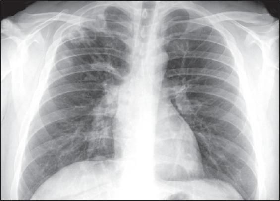
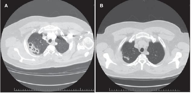
A chest radiograph revealed multiple cavitary lesions in the right upper lobe (Figure 1). A subsequent CT scan of the chest revealed multiple patchy areas of consolidation with cavitation most notable in the right lung (Figure 2A).

Figure 1 – Multiple cavitary lesions appear in the right upper lobe. This chest radiograph was obtained from a 26-year-old man who presented with cough and pleuritic chest pain.

Figure 2 – A CT scan of the chest revealed multiple patchy areas of consolidation with cavitation most notable in the right lung (A). After 4 weeks of fluconazole therapy, the patient's cough, hemoptysis, and pleuritic chest pain had resolved, and the CT scan documented significant improvement (B).
The patient was admitted to the general medicine floor with airborne precautions. Initial antibiotic therapy was withheld in the hopes of identifying a specific pathogen. The results of tuberculin skin testing and tests for HIV, Coccidioides antibody, proteinase 3 antibody, and myeloperoxidase antibody were negative.
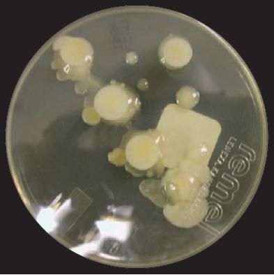
Because the patient's sputum and blood cultures were nondiagnostic, flexible fiberoptic bronchoscopy was performed. Bronchoalveolar lavage (BAL) of the right upper lobe revealed no organisms on direct examination; however, yeast grew on fungal culture (Figure 3). Yeast biochemical testing confirmed Cryptococcus neoformans as the pathogen.

Figure 3 – Cryptococcus neoformans grew on culture of a specimen obtained by bronchoalveolar lavage. The patient's sputum and blood cultures had been nondiagnostic.
A lumbar puncture was not performed because of the paucity of neurological signs and symptoms. The result of a serum Cryptococcus antigen test was positive, with a titer of 1:64. The patient was given oral fluconazole, 400 mg/d. He remained stable throughout his hospitalization, with resolution of his low-grade fever and a significant improvement in his cough and chest pain. He was discharged to home on a regimen of antifungal therapy.
After 4 weeks of fluconazole therapy, the patient's cough, hemoptysis, and pleuritic chest pain had resolved. A CT scan of the chestrevealed significant improvement (Figure 2B). Of interest, his Cryptococcus antigen titer had increased to 1:254 after 4 weeks of therapy.
Discussion
C neoformans is an encapsulated yeast that is found worldwide. It first became associated with pigeons when it was found in soil contaminated with pigeon excreta in the 1950s.1 Human exposure and infection are believed to occur via inhalation of aerosolized spores.
The vast majority of patients with cryptococcal pneumonia are immunocompromised, most commonly as a result of HIV infection, transplant-associated immunosuppression, or long-term corticosteroid use. In these patients, hematogenous spread, especially to the CNS, is common. Healthy persons rarely become infected with Cryptococcus and, when they do, dissemination and CNS infection are rare.
A small number of series have evaluated the differences between immunosuppressed and immunocompetent patients infected with C neoformans. Not only do these patients differ in presentation, site of infection, and rate of dissemination, but they also differ in treatmentapproaches and outcomes. Overall, immunocompetent patients with cryptococcosis do much better than immunocompromised patients. In fact, many immunocompetent patients improve without antifungal therapy.
Clinical presentation
The presenting clinical signs and symptoms of isolated pulmonary cryptococcosis vary in immunocompetent patients. The mean age of patients at presentation ranges from 41 to 61 years, without a significant difference between males and females.2,3 In one review, 13 immunocompetent patients with pulmonary cryptococcosis were compared with 16 immunocompromised patients, and the latter were found to be significantly older at diagnosis (41.2 years vs 59.8 years, respectively).2
The most common form of cryptococcal infection in immunocompetent patients is pneumonia. Approximately 31% to 50% of patients who have cryptococcal pneumonia are asymptomatic, and radiographic abnormalities are found incidentally.2-4 The most common symptom is cough, followed by dyspnea, chest pain, hemoptysis, and constitutional symptoms.
Radiographic findings
In immunocompetent patients who have pulmonary cryptococcosis, radiographic findings vary immensely. Nodules are the most common finding on plain chest radiographs, while bilateral nodules are frequently found on CT scans.5,6 The nodules may be cavitary, but this finding occurs more frequently in immunocompromised patients.2,7 Other rare findings on CT scans include ground-glass infiltrates, consolidation, lymphadenopathy, and pleural effusions.2,6
Serum Cryptococcus antigen
Methods of diagnosing pulmonary cryptococcosis include sputum culture, BAL, needle aspiration of pulmonary nodules or lymph nodes, transbronchial biopsy, and surgical resection. Serum Cryptococcus antigen titers can be used to document disease and for evidence of treatment response. Overall, titers seem to be higher on average in immunosuppressed patients than in immunocompetent patients.
In most studies, serum Cryptococcus antigen titers were decreased or were negative after treatment. 2,3,8-10 Of interest, our patient had an increased titer after 4 weeks of fluconazole therapy, despite improvement in symptoms and CT findings. The significance of the increase in serum Cryptococcus antigen titers is unknown.
There is no consensus on the role of Cryptococcus antigen testing or cerebrospinal fluid (CSF) culture in immunocompetent patients. Some experts advocate CSF analysis in all patients, while others test on the basis of symptoms. This latter approach appears to be reasonable, considering the very low rate of dissemination in immunocompetent patients, especially those who are asymptomatic.
In a study by Chang and associates,2 10 of 13 immunocompetent patients with pulmonary cryptococcosis had an initial positive serum antigen titer. A CSF culture was positive for Cryptococcus in 1 of 8 patients; it is unclear whether the patient with the positive CSF culture had any neurological clinical signs or symptoms. Antigen titers were decreased or negative at 12 months in all 9 patients for whom follow-up data were available. In 1 patient, the antigen titer initially rose despite antifungal therapy, but eventually it became undetectable.
Nadrous and associates3 reviewed 42 immunocompetent patients who had cryptococcosis diagnosed at their institution over 26 years; 36 of the patients had pulmonary involvement alone. Eleven patients underwent lumbar puncture, and in all, findings were negative. Of the 22 patients in whom serum Cryptococcus antigen was measured, only 3 had positive titers. Follow-up antigen titers were not reported in this study.
In a smaller series of 4 immunocompetent patients, all had positive serum antigen titers, and 3 of 4 had negative CSF cultures.4 Furthermore, all 4 had either negative or decreased serum antigen titers after antifungal treatment.
Treatment
No randomized controlled trials have studied the treatment of isolated pulmonary cryptococcosis in immunocompetent patients. At present, there is no clear consensus on treatment in this patient population. In the above-described reviews, resolution of disease occurred in 5 of 13 patients2 and in 8 of 17 patients3 who were not treated. Thus, it seems that despite the absence of therapy, symptomatic patients can still have disease resolution.
Mild, subclinical cryptococcal infections may be managed without the use of antifungal agents. For immunocompetent patients who have isolated pulmonary cryptococcosis, the decision not to treat is made more difficult by the availability of inexpensive, nontoxic antifungal medications. When amphotericin B was the only available therapy for cryptococcal pneumonia, many physicians did not treat these otherwise healthy patients because of the potential adverse effects of treatment and the often benign course of the disease.
However, there are reports of disseminated disease developing in immunocompetent patients with pulmonary infection. Chang and associates11 described an immunocompetent patient who had cryptococcal empyema and osteomyelitis of the adjacent rib. It has also been well documented that cryptococcal meningitis can occur in otherwise healthy persons.12,13
Emmons and associates14 described a healthy 46-year-old man who was found to have an incidental lingular mass on a routine chest radiograph. Surgical resection revealed a cryptococcoma. The serum Cryptococcus antigen titer was positive, but CSF findings were negative. Despite resection and treatment with fluconazole, 400 mg/d, the antigen titer continued to rise. Bronchoscopy revealed a new endobronchial mass, and cultures grew cryptococcal organisms. The patient was then treated with amphotericin B and 5-flucytosine for 6 weeks, with a decrease in the size of the mass and in serum antigen titers.
Treatment recommendations for cryptococcosis have been published by the Infectious Diseases Society of America (IDSA).15 Immunocompetent patients who have isolated pulmonary disease may be observed without therapy if CNS involvement has been excluded; however, the IDSA recommends treating all patients who are symptomatic. The recommended treatment is oral fluconazole (itraconazole is an acceptable alternative) for 3 to 6 months.15
Our decision to treat with antifungal therapy was based on the severity and duration of our patient's symptoms and on the extent of his parenchymal disease, which included a necrotizing pneumonia pattern. Our patient had an excellent response to treatment, with resolution of cough and pleuritic chest pain. The radiographic findings also demonstrated significant improvement of the pulmonary parenchymal abnormalities.
References:
REFERENCES
1.
Casadevall A, Perfect JR.
Cryptococcus neoformans.
Washington, DC: American Society for Microbiology; 1998.
2.
Chang WC, Tzao C, Hsu HH, et al. Pulmonary cryptococcosis: comparison of clinical and radiographic characteristics in immunocompetent and immunocompromised patients.
Chest.
2006;129:333-340.
3.
Nadrous HF, Antonios VS, Terrell CL, Ryu JH. Pulmonary cryptococcosis in nonimmunocompromised patients.
Chest.
2003;124:2143-2147.
4.
Núñez M, Peacock JE Jr, Chin R Jr. Pulmonary cryptococcosis in the immunocompetent host. Therapy with oral fluconazole: a report of four cases and a review of the literature.
Chest.
2000;118:527-534.
5.
Khoury MB, Godwin JD, Ravin CE, et al. Thoracic cryptococcosis: immunologic competence and radiologic appearance.
AJR.
1984;142:893-896.
6.
Lindell RM, Hartman TE, Nadrous HF, Ryu JH. Pulmonary cryptococcosis: CT findings in immunocompetent patients.
Radiology.
2005;236:326-331.
7.
Kishi K, Homma S, Kurosaki A, et al. Clinical features and high-resolution CT findings of pulmonary cryptococcosis in non-AIDS patients.
Respir Med.
2006;100:807-812.
8.
Yamaguchi H, Ikemoto H, Watanabe K, et al. Fluconazole monotherapy for cryptococcosis in non-AIDS patients.
Eur J Clin Microbiol Infect Dis.
1996;15:787-792.
9.
Nakamura S, Miyazaki Y, Higashiyama Y, et al. Community acquired pneumonia (CAP) caused by
Cryptococcus neoformans
in a healthy individual.
Scand J Infect Dis.
2005;37:932-935.
10.
Aberg JA, Watson J, Segal M, Chang LW. Clinical utility of monitoring serum cryptococcal antigen (sCRAG) titers in patients with AIDSrelated cryptococcal disease.
HIV Clin Trials.
2000;1:1-6.
11.
Chang WC, Tzao C, Hsu HH, et al. Isolated cryptococcal thoracic empyema with osteomyelitis of the rib in an immunocompetent host.
J Infect.
2005;51:e117-e119.
12.
Lui G, Lee N, Ip M, et al. Cryptococcosis in apparently immunocompetent patients.
QJM.
2006;99:143-151.
13.
Reichman N, Elias M, Raz R, Flatau E. Cryptococcal meningitis following cryptococcal pneumonia in an immunocompetent [in Hebrew].
Harefuah.
1999;137(7-8):291-292, 350.
14.
Emmons WW 3rd, Luchsinger S, Miller L. Progressive pulmonary cryptococcosis in a patient who is immunocompetent.
South Med J.
1995;88:657-660.
15.
Saag MS, Graybill RJ, Larsen RA, et al. Practice guidelines for the management of cryptococcal disease. Infectious Diseases Society of America.
Clin Infect Dis.
2000;30:710-718.
