- Clinical Technology
- Adult Immunization
- Hepatology
- Pediatric Immunization
- Screening
- Psychiatry
- Allergy
- Women's Health
- Cardiology
- Pediatrics
- Dermatology
- Endocrinology
- Pain Management
- Gastroenterology
- Infectious Disease
- Obesity Medicine
- Rheumatology
- Nephrology
- Neurology
- Pulmonology
Contact Dermatitis to Poison Ivy and Photoallergic Reaction to NSAID
Dermclinic

Case 1:
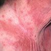
This pruritic eruption developed on a 62-year-old man's neck and upper chest after he had mowed his lawn. He has not started any new medications, and he has no recent history of contactant exposure. He did not mow any new sections of his yard.
Which of the following do you suspect?
A.
Contact dermatitis to a poisonous plant, such as poison ivy.
B.
Contact dermatitis to sunscreen.
C.
Photoallergic reaction to medication.
D.
Irritation (chafing) from heat and humidity.
E.
Contact dermatitis to cologne.
Bonus question: What clinical clue here points to the diagnosis?
(Answer on next page.)

Case 1: Contact dermatitis to a poisonous plant
This patient had an allergic reaction to a poisonous plant, such as poison ivy, A. The distribution of the rash corresponds to areas that were exposed to the lawn clippings from mowing. Cologne can produce a similar reaction.
Reactions to sunscreen can occur, although they are rare and would be present on all areas to which sunscreen had been applied. The 2 types of sunscreen reactions are a simple contact dermatitis to the product and a photo- allergic reaction on sun-exposed areas to which sunscreen had been applied.
A photoallergic reaction to medication would occur on all sun-exposed skin. Sparing of skin that has not been exposed to sunlight helps distinguish photo-aggravated conditions from contact dermatitis.
Finally, heat and humidity usually produce a rash in the groin region and between folds of skin, rather than on the neck.
Answer to bonus question: The linear streak on the side of the neck suggests an external contact dermatitis.

Case 2:
A 63-year-old man seeks evaluation of a worsening pruritic rash that appeared on his neck, upper chest, and lower half of his face 1 day earlier. He has been taking glucosamine/chondroitin and a lipid-lowering agent for several years, and he recently started taking an NSAID intermittently for mild arthritis.
What is the most likely cause of the rash?
A.
Contact dermatitis to a poisonous plant, such as poison ivy.
B.
Contact dermatitis to sunscreen.
C.
Photoallergic reaction to medication.
D.
Irritation (chafing) from heat and humidity.
E.
Contact dermatitis to cologne.
(Answer on next page.)

Case 2: Photoallergic reaction to NSAID
The NSAID that this patient had recently started taking caused a photoallergic reaction, C. He was advised to use a UVA-blocking sunscreen with a high sun-protection factor if he wished to continue NSAID therapy or to switch to an alternative medication that was not photosensitizing.
Contact dermatitis to a poisonous plant or cologne would be possible if there was a history of exposure. Irritation from heat and humidity is less likely because it typically causes a rash in the inguinal and intertriginous regions.

Case 3:
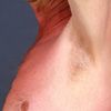
A 34-year-old woman has an area of asymptomatic hyperpigmentation on her shoulder; it has been present since birth. The use of sunscreen has no effect on the lesion.
Do you recognize this lesion?
A.
Giant café-au-lait spot.
B.
Mongolian spot.
C.
Nevus of Ito.
D.
Futcher lines.
E.
Becker nevus.
(Answer on next page.)

Case 3: Nevus of Ito
This patient has a nevus of Ito, C, on her shoulder. This congenital lesion is typically in the distribution of the posterior supraclavicular and lateral cutaneous brachial nerves. Although most nevi of Ito are present at birth, some appear at puberty.
Becker nevi also appear at puberty; however, terminal hairs are usually present within the lesion. Mongolian spots resemble nevi of Ito, but they typically disappear during childhood. Futcher lines are melanocyte migration lines that are more commonly seen on the upper arm of persons with black skin. Giant café-au-lait spots are usually well demarcated, unlike this patient's lesion.

Case 4:
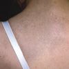
For several years, a 49-year-old woman has had persistent pruritus just lateral to the midline of her mid-back. She is unaware of any exacerbating factors. She has tried wearing a different type of bra, but her symptoms have not abated.
What is your clinical impression?
A.
Café-au-lait spot.
B.
Tinea versicolor.
C.
Contact dermatitis.
D.
Notalgia paresthetica.
E.
Fixed drug eruption.
(Answer on next page.)

Case 4: Notalgia paresthetica
Notalgia paresthetica, D, is characterized by pruritus and/or a burning sensation on the mid-back; a pigmented patch is frequently found in the involved area. This unilateral sensory neuropathy is often caused by degenerative changes in the corresponding vertebrae, which lead to spinal nerve impingement. Treatment options include topical anti-pruritic agents (not corticosteroids), paravertebral blocks, and lidocaine patches.
Café-au-lait spots are well demarcated and asymptomatic. Contact dermatitis and fixed drug eruptions feature erythema and scale, which are not present here. Tinea versicolor usually consists of discrete scaling macules.
