- Clinical Technology
- Adult Immunization
- Hepatology
- Pediatric Immunization
- Screening
- Psychiatry
- Allergy
- Women's Health
- Cardiology
- Pediatrics
- Dermatology
- Endocrinology
- Pain Management
- Gastroenterology
- Infectious Disease
- Obesity Medicine
- Rheumatology
- Nephrology
- Neurology
- Pulmonology
Compound Fracture of the Wrist
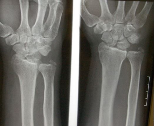
Figure 1. Initial x-ray of left wrist injury. (Click to enlarge)

Figure 2. (Click to enlarge)

A 52-year-old man presents to the emergency department with a left wrist injury after a ground level fall. He states that he felt fine before slipping on a wet floor in his kitchen. There was no loss of consciousness, weakness, dizziness, chest pain, or preceding illness and he denies palpitations. He states that he did not hit his head and reports no neck or back pain or other injuries. He has no other complaints. There is no significant past medical history and there are no known risk factors for osteoporosis although he does smoke cigarettes.
On physical examination, the patient’s vital signs are normal and he appears to be in mild to moderate distress. Examination of the head, neck, back, and torso are all unremarkable. Examination of the extremities shows swelling and tenderness diffusely about the left wrist with intact pulses and sensation, intact skin, and no tense compartments. The elbow and hand are not tender.
His wrist x-ray fim is shown in Figure 1.
What injury or injuries can you identify?
Answer: There are 4 injuries
The x-ray films demonstrated the following injuries: ulnar styloid fracture, “die-punch” intra-articular distal radius fracture, scaphoid fracture, and finally, an anterior lunate dislocation (Figure 2). Of these, the latter two are the most difficult to see.
Discussion
The ulnar styloid fracture is displaced and easily visualized on the previous views. The distal radius fracture is non-displaced and only involves the outer side of the distal radius. It is a type of die-punch fracture caused by the scaphoid bone being driven directly into the distal radius. With this type of fracture the scaphoid is also often injured.
The scaphoid bone, which sits directly distal to the radius on the radial side of the wrist, also is damaged with a non-displaced fracture at its waist or mid-portion. It is important to be aware of the fact that scaphoid fractures are often radiographically occult and so if there is snuff box tenderness on physical exam or any other exam findings that suggest a scaphoid injury, the wrist should be splinted and the patient treated as if there were a fracture regardless of plain film findings. For a potential scaphoid fracture the patient should be referred to orthopedics for definitive management, which may include delayed radiography and/or MRI, the latter being more sensitive than plain films.
The final injury is an anterior lunate dislocation, which is difficult to see except on the lateral view, which was not included. The complete wrist series (Figure 2) shows the lunate bone tipped forward to the left. Note that in a lateral view of a normal wrist the proximal capitate bone sits in the “cup” of the lunate, but in the injured wrist this alignment is lost. (See all the bones of the wrist.)
Outcome
This patient was actually splinted for a wrist fracture by the initial provider, with the latter two injuries having been missed. He returned the next day because the pain was not relieved by hydrocodone, and the prior day’s x-ray films were reviewed. The lunate dislocation was reduced with the assistance of orthopedics, and the pain was much improved. Fortunately there was no median nerve injury. The fractures healed well with 6 weeks of casting.
