- Clinical Technology
- Adult Immunization
- Hepatology
- Pediatric Immunization
- Screening
- Psychiatry
- Allergy
- Women's Health
- Cardiology
- Pediatrics
- Dermatology
- Endocrinology
- Pain Management
- Gastroenterology
- Infectious Disease
- Obesity Medicine
- Rheumatology
- Nephrology
- Neurology
- Pulmonology
Common Shoulder Problems:
Our goal here is to help you master the shoulder examination. We review the basics of the examination, and we evaluate emerging concepts in the diagnosis of the more common shoulder conditions.
Clinicians find the shoulder difficult to examine for several reasons:
- It has multiple complex parts, including the glenohumeral joint, the acromioclavicular joint, the sternoclavicular joint, and the scapulothoracic articulation.
- Many of the pathologic processes that affect the shoulder are poorly understood. For example, although rotator cuff disease has been thought to result from impingement of the rotator cuff on the acromion, many studies now suggest that senescence of the tendon is a more important factor and that impingement against bone spurs may not play as large a role.1-3 In addition, pain elicited during a shoulder examination that once was thought to be caused by tendons hitting spurs may instead result from tendons impinging against the superior glenoid rim.1,2,4
- Access to the affected area is restricted because of soft tissue coverage of the glenohumeral joint (by the deltoid and other muscles). Although palpation of the acromioclavicular joint is relatively easy, accurate palpation of rotator cuff tears or the biceps tendon is nearly impossible.
- Pain patterns related to various conditions can overlap and not be specific for any one entity. For example, although pain in the deltoid or proximal humerus may result from rotator cuff problems, it also may be caused by stiffness, cervical radiculopathy, arthritis, and numerous other conditions.
- Because many shoulder tests are sensitive for detecting abnormalities but are not specific for any one condition, they have less diagnostic value than was once thought (Table).5-7
- A thorough evaluation often is needed to narrow the differential diagnosis and determine proper treatment, because more than one condition may be causing the patient's symptoms.
Our goal here is to help you master the shoulder examination. We review the basics of the examination, and we evaluate emerging concepts in the diagnosis of the more common shoulder conditions.
GENERAL PRINCIPLES OF EVALUATION
Key factors in the history. A major factor in examination, evaluation, and management of shoulder problems is the timing of symptom onset (acute with a history of trauma or insidious).7-9 For symptoms of acute or traumatic onset, the differential diagnosis includes fractures, acromioclavicular joint injuries, biceps tendon injuries, rotator cuff tears, dislocations, and subluxations. For symptoms of insidious onset, the differential diagnosis typically includes rotator cuff problems; stiff, or frozen, shoulder; arthritis of any cause; cancer; and cervical radiculopathy. If a patient's symptoms have begun after participation in a new exercise or activity without trauma, an exacerbation of preexisting rotator cuff problems or joint arthritis has probably occurred; a subsequent examination can help determine the cause.
The next important factor in the history is the nature of the patient's complaint (pain, weakness, or loss of motion). Painless weakness of insidious onset is a neurologic problem until proved otherwise. Acute painless weakness after a traumatic event also should alert the examiner to a possible neurologic cause, but if the trauma site was distant from the shoulder, rotator cuff tears may present as painless weakness.
Painful weakness after a traumatic event may have multiple causes, each of which needs to be ruled out with a careful examination and ancillary studies. Painful, insidious weakness may be the result of cervical radiculopathy, chronic rotator cuff problems, tumors, or arthritis. Traumatic loss of range of motion may indicate a fracture or a torn rotator cuff. Insidious loss of range of motion may be caused by weakness or shoulder stiffness.
Physical examination basics. Addressing the patient's complaint can help you determine the cause of the shoulder problem by directing the examination and subsequent studies. For example, the patient who complains of loss of motion should be examined to determine whether the active and passive range of motion in the affected shoulder are the same or different.

If they are the same, any process that tightens up the joint (eg, adhesive capsulitis or arthritis of any cause) should be considered. If active motion is less than passive motion in a patient who has no pain, a neurologic process or a torn rotator cuff should be suspected. If a patient complains of weakness, loss of motion, and paresthesias, especially in the absence of trauma, a neurologic cause should be ruled out.
Axioms for shoulder examination that may improve your ability to accurately determine the cause of the patient's complaints include the following:
- Ask the patient to undress so that both shoulders can be examined from the front and the back. Atrophy of the muscles, especially the supraspinatus and infraspinatus, is a clue to a neurologic or rotator cuff disorder (Figure 1).
- Compare the affected and unaffected shoulders because atrophy and deformity may be subtle in patients who have shoulder complaints.
- A neurologic evaluation of the upper extremity should always be part of the initial examination. Include a brief sensory examination of the whole upper extremity, range of motion assessment, and strength testing. Among the motions the patient should perform is full elevation of the shoulder in flexion or abduction, reaching to the top of the head and moving a hand as far up the back as possible. Loss of motion or asymmetry of motion indicates weakness or stiffness.
- Imaging studies should always begin with plain radiographs (in 2 planes) because they may preclude the need for other studies, such as MRI. Obtain an anteroposterior view of the shoulder in internal rotation and a true anteroposterior (Grashey) view with the arm in external rotation; the latter best shows the joint space of the glenohumeral joint. The axillary view is the best for evaluating the shoulder in a second plane; in cases of acute trauma, a scapular Y view may suffice.10
EVALUATING SPECIFIC SHOULDER PROBLEMS
Rotator cuff abnormalities. Rotator cuff degeneration is a normal consequence of aging. Tendinitis of the rotator cuff will occur in most persons at least once in their lives.


There are 2 ways to tear the rotator cuff: trauma and an attritional process. The latter is similar to "wearing a hole in the seat of your pants"; the tear occurs gradually and insidiously without the patient being aware of it. Partial and even full-thickness tearing of the rotator cuff as a result of this insidious wear is common, and the incidence increases with age.11-13
Many of these rotator cuff tears are asymptomatic and do not require management. A patient with insidious onset of pain who is found to have a rotator cuff tear (either partial or full) probably aggravated a preexisting abnormal (or torn) rotator cuff tendon.
The rotator cuff is not needed for good shoulder range of motion. One study has shown that anesthetizing the suprascapular nerve, which innervates the supraspinatus and infraspinatus muscles, does not preclude full arm elevation.14 The main finding in patients who have a full-thickness rotator cuff tear is weakness in abduction and in external rotation. However, even patients with long-standing rotator cuff disease who have learned to compensate by using the deltoid muscle may have full range of motion and may show only mild to moderate weakness on examination.
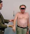
Therefore, as recent studies have shown, the most important tests in the examination of patients for rotator cuff problems are those of strength and motion.5,15 The drop arm test, one of the most diagnostic tests for a full-thickness rotator cuff tear,16 is performed in 1 of 2 ways: ask the patient to raise his or her arm to full elevation and then to bring the arm down slowly (if he cannot maintain this slow descent against gravity, he may have a rotator cuff tear or weakness of any cause) or ask the patient to elevate the arm to 90 degrees and then hold it there (Figures 2 and 3). If the patient cannot maintain that elevation, consider the diagnosis to be a rotator cuff tear or weakness of any cause.


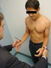
Weakness, especially in external rotation or abduction, is another important physical finding that indicates full-thickness rotator cuff tears. Testing of strength in external rotation is accomplished by having the patient place his arms at his sides with the elbows bent 90 degrees. The examiner pushes against the patient's wrists (Figure 4). Weakness in the patient's resistance indicates some process affecting the infraspinatus muscle. However, such weakness may result from pain alone, cervical disk disease, or injury to the infraspinatus nerve by synovial cysts. Studies have shown this test to be sensitive, but not specific, for rotator cuff tears.5
In the past, it was suggested that testing for supraspinatus strength be performed in the "empty can" position (the patient's arms abducted 90 degrees, elbows extended, and arms internally rotated) (Figure 5).9 However, it now appears that this test is equally effective with the arms in neutral or with the thumbs up (the "full can" position).17 This finding is important because the thumbs-down position tends to be painful and may provide false-positive results caused by pain. In addition, the supraspinatus strength test assesses not only the supraspinatus but also the deltoid muscle.17 As a result, some patients with a strong deltoid muscle may have good strength with resisted abduction despite having a torn rotator cuff.
Other popular tests for the evaluation of rotator cuff abnormalities are the impingement signs. In the Neer sign examination, the examiner elevates the arm of the standing patient into flexion (Figure 6 – Please see CONSULTANT, April 15, 2005, page 444).18,19 Pain elicited in the deltoid is a positive test result. This test has a sensitivity of 68% and a specificity of 68.7% for rotator cuff abnormalities of any type (tendinitis alone, partial rotator cuff tears, or full-thickness rotator cuff tears), but the result may be positive because of a variety of shoulder conditions.5,18,19
In the Hawkins-Kennedy test, the examiner elevates the patient's affected shoulder to 90 degrees and internally rotates the arm.20 Pain in the deltoid is a positive test result. The positive result may be seen in patients with a stiff shoulder from any cause; in patients with a stiff shoulder, the pain frequently is posterior because this maneuver stretches the posterior shoulder. Like the Neer impingement sign, this test is better for low grades of rotator cuff abnormalities, such as painful tendinosis and partial rotator cuff tears.5
Combining tests greatly increases your ability to diagnose a full-thickness rotator cuff tear.5,15 Park and associates5 found that if a patient has a positive drop arm test result, a painful arc, and weakness in external rotation, there is a greater than 90% chance that he has a full-thickness rotator cuff tear. Murrell and Walton15 found that if a patient has weakness in abduction and in external rotation, as well as a positive impingement sign, there is a 98% chance that he has a full-thickness rotator cuff tear.
Stiff shoulder. In our practice, the condition most frequently mistaken for rotator cuff problems is a stiff shoulder. Both conditions may begin insidiously without trauma and can be very painful. To complicate matters, a stiff shoulder can coexist with rotator cuff problems or other causes of shoulder pain.
A patient with rotator cuff problems typically complains of pain with motions over shoulder level or when lifting away from the body; a patient with stiff shoulder does not have pain until the limit of the motion (limit of capsule length) is reached ("end-point pain"). A patient with frozen shoulder often complains of difficulty in tucking in a shirt or fastening a bra or of the inability to put on a coat, reach behind, or swipe a card reader in a parking garage. On examination, a Hawkins-Kennedy impingement sign often is positive because of the loss of internal rotation.
When you examine a patient who has a painful shoulder, compare the affected and unaffected sides. Also, when a patient reaches the limit of active motion, gently try to establish whether more passive motion is possible. If it is, the cause of motion loss may be weakness or a rotator cuff tear. If no more motion is possible, the patient has stiff shoulder by definition.
A subacromial injection may be used to distinguish the pain of a stiff shoulder from that of a rotator cuff problem. If an injection of local anesthetic into the subacromial space provides relief within a few minutes, the most likely diagnosis is bursitis associated with rotator cuff problems. If it provides no immediate relief (within 5 to 10 minutes) and the patient's shoulder is stiff, the cause of the pain probably is stiffness rather than rotator cuff tendinitis.
Acromioclavicular joint conditions. These may be divided into those of traumatic or nontraumatic origins. Traumatic origins include a fall directly onto the shoulder and a blow delivered directly to the point of the shoulder, such as is seen in hockey players who are checked into the boards by opposing players. To rule out fracture, obtain x-ray films for patients who have a history of falling and landing on the shoulder and who have distal clavicle tenderness or a deformity. Acromioclavicular joint sprains produce swelling and, often, deformity; the diagnosis may be confirmed with radiography.
Nontraumatic origins include osteolysis of the distal clavicle (caused by bench-pressing weights or performing push-ups) and chronic stress on the acromioclavicular joint resulting from repetitive activity. Chronic conditions of the acromioclavicular joint may be diagnosed by detecting local tenderness. Acromioclavicular joint pain may radiate into the neck and simulate cervical disk problems. Injection of local anesthetic into this joint may confirm the diagnosis.
Other tests for acromioclavicular joint abnormalities include the cross-body adduction stress test; the active compression, or O'Brien, test21; and the arm extension test.6 The accuracy of these tests is variable; the most important diagnostic finding for symptomatic acromioclavicular joint abnormality is tenderness to palpation over the joint.
Acromioclavicular arthritis, which may begin in the second decade of life, increases linearly with age22 and often is asymptomatic. One study of MRI scans of asymptomatic shoulders showed a greater than 90% incidence of acromioclavicular arthritis in persons older than 30 years.22 Therefore, acromioclavicular arthritis is almost a normal variant. The diagnosis of symptomatic acromioclavicular arthrosis should be based on detection of local tenderness or on results of other supporting diagnostic tests.
References:
REFERENCES:
1.
McCallister WV, Parsons IM, Titelman RM, Matsen FA 3rd. Open rotator cuff repair without acromioplasty.
J Bone Joint Surg Am
. 2005;87:1278-1283.
2.
Voloshin I, Gelinas J, Maloney MD, et al. Proin-flammatory cytokines and metalloproteases are ex-pressed in the subacromial bursa in patients with rotator cuff disease.
Arthroscopy.
2005;21:1076.
3.
Yuan J, Wang MX, Murrell GA. Cell death and tendinopathy.
Clin Sports Med
. 2003;22:693-701.
4.
Budoff JE, Rodin D, Ochiai D, Nirschl RP. Arthroscopic rotator cuff debridement without decompression for the treatment of tendinosis.
Arthroscopy.
2005;21:1081-1089.
5.
Park HB, Yokota A, Gill HS, et al. Diagnostic ac-curacy of clinical tests for the different degrees of subacromial impingement syndrome.
J Bone Joint Surg
. 2005;87A:1446-1455.
6.
Chronopoulos E, Kim TK, Park HB, et al. Diagnostic value of physical tests for isolated chronic acromioclavicular lesions.
Am J Sports Med.
2004;32: 655-661.
7.
McFarland EG, Kim TK.
Examination of the Shoulder: The Complete Guide.
New York: Thieme. In press.
8.
Krishnan SG, Hawkins RJ, Bokor DJ. Clinical evaluation of shoulder problems. In: Rockwood CA Jr, Matsen FA 3rd, Wirth MA, Lippitt SB, eds.
The Shoulder
. 3rd ed. Philadelphia: WB Saunders; 2004: 145-185.
9.
McFarland EG, Shaffer B, Glousman RE, et al. Anterior shoulder instability, impingement, and rotator cuff tear. Section B: Clinical and diagnostic evaluation. In: Jobe FW, ed.
Operative Techniques in Upper Extremity Sports Injuries
. St Louis: Mosby-Year Book Inc; 1996:177-190.
10.
Harvey RA, Trabulsy ME, Roe L. Are postreduction anteroposterior and scapular Y views useful in anterior shoulder dislocations?
Am J Emerg Med.
1992;10:149-151.
11.
Brewer BJ. Aging of the rotator cuff.
Am J Sports Med
. 1979;7:102-110.
12.
Milgrom C, Schaffler M, Gilbert S, Van Holsbeeck M. Rotator-cuff changes in asymptomatic adults. The effect of age, hand dominance and gender.
J Bone Joint Surg Br
. 1995;77:296-298.
13.
Tempelhof S, Rupp S, Seil R. Age-related prevalence of rotator cuff tears in asymptomatic shoulders.
J Shoulder Elbow Surg
. 1999;8:296-299.
14.
Colachis SC Jr, Strohm BR. Effect of suprascapular and axillary nerve blocks on muscle force in upper extremity.
Arch Phys Med Rehabil
. 1971;52:22-29.
15.
Murrell GA, Walton JR. Diagnosis of rotator cufftears [published correction appears in
Lancet
. 2001;357:1452].
Lancet.
2001;357:769-770.
16.
Codman EA. Rupture of the supraspinatus ten-don. In:
The Shoulder: Rupture of the Supraspinatus
Tendon and Other Lesions in or About the Subacromial Bursa.
Boston: Thomas Todd Co; 1934:123-177.
17.
Kelly BT, Kadrmas WR, Speer KP. The manual muscle examination for rotator cuff strength. An electromyographic investigation.
Am J Sports Med
. 1996;24:581-588.
18.
Neer CS 2nd. Anterior acromioplasty for the chronic impingement syndrome in the shoulder: a preliminary report.
J Bone Joint Surg Am
. 1972;54: 41-50.
19.
Neer CS 2nd. Impingement lesions.
Clin Orthop Relat Res
. 1983;173:70-77.
20.
Hawkins RJ, Kennedy JC. Impingement syndrome in athletes.
Am J Sports Med
. 1980;8:151-158.
21.
O'Brien SJ, Pagnani MJ, Fealy S, et al. The active compression test: a new and effective test for diagnosing labral tears and acromioclavicular joint abnormality.
Am J Sports Med.
1998;26:610-613.
22.
Stein BE, Wiater JM, Pfaff HC, et al. Detection of acromioclavicular joint pathology in asymptomatic shoulders with magnetic resonance imaging
. J Shoulder Elbow Surg
. 2001;10:204-208.
