- Clinical Technology
- Adult Immunization
- Hepatology
- Pediatric Immunization
- Screening
- Psychiatry
- Allergy
- Women's Health
- Cardiology
- Pediatrics
- Dermatology
- Endocrinology
- Pain Management
- Gastroenterology
- Infectious Disease
- Obesity Medicine
- Rheumatology
- Nephrology
- Neurology
- Pulmonology
Chronic Venous Disease: Evaluation of the Various Manifestations
A description of the evaluation of the various manifestations of chronic venous disease.
Venous disease has been recognized for more than 3000 years. Varicose veins were described in an Egyptian papyrus and depicted at the base of the Acropolis.1
In the United States, at least 25 million persons have varicosities, 2 to 6 million have advanced changes of chronic venous insufficiency (edema, skin changes), and about 500,000 (14% of persons older than 65 years) have venous ulcers.2 In addition, 10% to 35% of American adults have some form of chronic venous disorder, ranging from spider veins and simple varicosities to the most advanced form of chronic venous insufficiency-ulcers.3,4 The associated economic consequences-time off from work (estimated at 4.6 million work days per year), disability, and early retirement-total billions of dollars. The cost to the government for treatment of advanced venous disease and venous ulcer care exceeds 1 billion dollars.5
Here I describe the evaluation of the various manifestations of chronic venous disease. In a second article, I will discuss the options for managing both uncomplicated and advanced venous conditions.
ANATOMY
The venous system in the legs can be categorized as follows:
- The superficial system consists of the great saphenous vein, the small saphenous vein, and their branches.
- The deep system lies in the intermuscular portion of the leg and is responsible for more than 80% of the venous return from the leg.
- The perforator veins form a communication network between the superficial and deep veins, with flow normally directed from the superficial to the deep.
PATHOPHYSIOLOGY
Venous disease typically affects the legs because they are vulnerable to the hydrostatic pressures of gravity and the hydrodynamic pressure of the "calf pump," which can exceed 150 mm Hg.6 In a normal leg, all the veins have unidirectional bicuspid valves that keep blood flowing in an ascending direction. Problems arise when these valves malfunction and allow retrograde blood flow. The resultant increase in hydrostatic pressure is especially significant in the lower calf and malleolar region.
Elevated hydrostatic pressure leads to a transcapillary passage of fluid that contains proteins, red cells, and white cells. This fluid can cause inflammation of the pericapillary tissue with fibrin deposition7 and white cell-mediated tissue destruction.8 Over years, the tissue progressively deteriorates; this phenomenon is known as lipodermatosclerosis-a fibrotic destruction of the subcutaneous fat and the skin.
Patients with chronic venous insufficiency have ambulatory hypertension in their leg veins. This may result from primary valvular incompetence in superficial, deep, or perforator veins, or it may be secondary to vein wall injury from previous deep venous thrombosis. The inflammatory process related to the deep venous thrombosis generally leads to progressive venous valvular fibrosis and eventually to valvular insufficiency. This venous valvular insufficiency results in venous reflux, which is the hallmark of most cases of venous hypertension. Less commonly, venous hypertension is caused by persistent venous obstruction related to deep venous thrombosis that has not recanalized.
CLINICAL PRESENTATION
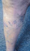
Spider veins. Telangiectases and blue, or reticular, veins are generally considered cosmetic problems (Figure 1). They can be associated with mild discomfort in the legs or localized discomfort in a particular pattern.
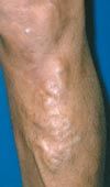
Varicose veins. Most patients who seek treatment have primary varicose veins without advanced sequelae (Figure 2). These patients typically complain of tiredness, aching, and throbbing of the involved extremities that worsen through the day. Complications of varicosity include thrombophlebitis, a slight predisposition to deep venous thrombosis, occasional bleeding from ruptured varices, swelling, and ulceration.
The most important risk factor for varicose veins is a positive family history. Evidence shows that varicosity is linked to an autosomal dominant gene with variable penetrance.9 Other risk factors are pregnancy, female gender, older age, obesity, and prolonged standing.
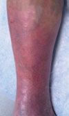
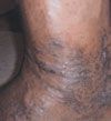
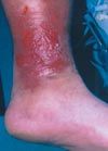

Chronic venous insufficiency. This is characterized by edema, lipodermatosclerosis, stasis dermatitis, and healed or active venous ulcerations. In patients with lipodermatosclerosis, the skin may initially have a pinkish hue (Figure 3), which becomes progressively darker. Eventually, a brownish discoloration develops (Figure 4). Stasis dermatitis, or stasis eczema, is an exacerbation of this process (Figure 5). This inflammatory reaction must be distinguished from cellulitis, because antibiotics are not effective in treating stasis dermatitis.
Skin changes with ulceration are more common in older patients and in those who have involvement of the deep system or perforator veins (Figure 6). About 80% of patients with venous ulcers have superficial venous incompetence in addition to deep or perforator vein incompetence. In a study of patients with venous ulcerations, duplex ultrasonography revealed isolated superficial vein incompetence in only 17% of patients and perforator vein incompetence in 63% of patients.10 Tissue changes are much more advanced in patients with previous deep venous thrombosis than in those with primary valvular incompetence.
CLASSIFICATION OF CHRONIC VENOUS DISEASE
The Clinical, Etiologic, Anatomic, and Pathologic (CEAP) classification was created to standardize the diagnosis of chronic venous disease. The disease is classified by the following11:
Clinical class. The Table lists the 7 clinical classes of chronic venous disease.
Etiology. A primary cause is undetermined; a secondary cause is known, such as previous thrombosis or trauma:
- Congenital (Ec).
- Primary (Ep).
- Secondary (Es).
Anatomic distribution. More than 1 venous system may be affected:
- Superficial (As).
- Perforator (Ap).
- Deep (Ad).
Pathology. Note that patients may have both obstruction and reflux in the same deep venous system:
- Reflux (Pr).
- Obstruction (Po).
- Reflux and obstruction (Pro).
DIAGNOSTIC TESTING
A careful history taking is essential before the venous evaluation. Include the following questions:
- Does the patient have a history of deep venous thrombosis?
- How long has the patient had swelling?
- How long have skin changes (eg, pigmentation) been present?
A thorough evaluation of the leg with duplex ultrasonography is a key component of the workup. This study is performed with the patient standing. Careful assessment of perforator veins is essential because incompetence of these veins is a significant cause of recurrence of varicose veins and can produce distal skin changes. The deep system must also be carefully evaluated for obstruction and incompetence. When chronic venous insufficiency is a concern, it is important to specify that the technician assess all 3 venous systems for valvular insufficiency.
Physiologic and radiographic tests can be helpful; however, they are generally not cost-effective. In most centers, physiologic tests, such as photoplethysmography and impedance plethysmography, along with ambulatory venous pressure measurements, have been replaced by duplex ultrasonography. Contrast phlebography is rarely used in the diagnosis or treatment of chronic venous insufficiency. Duplex ultrasonography provides the most accurate means of identifying sites of incompetence and differentiating deep from superficial or perforator reflux.
Obesity Linked to Faster Alzheimer Disease Progression in Longitudinal Blood Biomarker Analysis
December 2nd 2025Biomarker trajectories over 5 years in study participants with AD show steeper rises in pTau217, NfL, and amyloid burden among those with obesity, highlighting risk factor relevance.
