- Clinical Technology
- Adult Immunization
- Hepatology
- Pediatric Immunization
- Screening
- Psychiatry
- Allergy
- Women's Health
- Cardiology
- Pediatrics
- Dermatology
- Endocrinology
- Pain Management
- Gastroenterology
- Infectious Disease
- Obesity Medicine
- Rheumatology
- Nephrology
- Neurology
- Pulmonology
Chondrodermatitis Nodularis Helicis and Verruca Vulgaris
Skin lesions in the outer ear-the pinna, the concha, and the external auditory meatus-may be trivial or potentially malignant. Here we consider a flesh-colored swelling with a central white spicule; a tan papillary lesion; a fluctuant swelling; a red berry-like papule; an elevated pink lesion.
Test your diagnostic prowess on the outer-ear lesions at right:
Image 1: Flesh-colored swelling with a 1-mm white scab
Image 2: Tan, 7x3x5-mm, papillary lesion with a broad, slightly elevated, eccentric base
Image 3: Asymptomatic swelling in the superior crus of the right antihelix
Image 4: 0.5-cm, red berry-like lesion in the concha
Image 5: Light tan lesion with brown specks that is slightly raised and finely roughened
Image 6: Raised, elongated, light tan lesion on the anterior aspect of the superior helix
Image 7: Raised, pink lesion with a tiny scab on the anterior ridge of the left lower helix
(For further details, see the next page.)
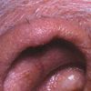
Case 1:
An 80-year-old man has had an asymptomatic, flesh-colored swelling on his right ear for 4 to 5 months. In the center is a 1-mm white scab pointing downward from the helix. At times, the patient shaves a white spicule that grows in this crusted area. He sleeps on his right side and does not use a cell phone.
What type of lesion is this, and how is it best removed? (Click here to find the answer.)
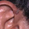
Case 2:
This tan, 7x3x5-mm, papillary lesion with a broad, slightly elevated, eccentric base has been present for several months on the right anterior helix of a 24-year-old man. It is asymptomatic.
What are you looking at here? (Click here to find the answer.)
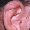
Case 3:
A 35-year-old woman is concerned about a 1-cm swelling that suddenly appeared in the superior crus of the right antihelix. The lesion is fluctuant and asymptomatic; no tenderness or warmth is felt on palpation. She denies trauma to the area.
How would you proceed? (Click here to find the answer.)
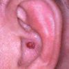
Case 4:
This asymptomatic, 0.5-cm, red berry-like lesion in the concha of a 39-year-old man's left ear has been gradually enlarging for 3 months.
Is this lesion cause for alarm? (Click here to find the answer.)
Figure A Figure B
Case 5:
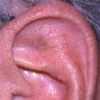
For several months, a 67-year-old man has had an asymptomatic, 0.5-cm, light tan lesion with brown specks on his left ear that is slightly raised and finely roughened (A).
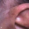
A 69-year-old woman presents with a raised, elongated, light tan lesion on the anterior aspect of the superior helix of about 1 month's duration (B). It is also asymptomatic.
Can you identify these lesions? (Click here to find the answer.)
Case 6:
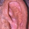
An 84-year-old man presents for evaluation of a raised, pink lesion with a tiny scab on the anterior ridge of the left lower helix. This asymptomatic, 0.5-cm lesion has been present for 2 months.
What approach would you take? (Click here to find the answer.)
References:
REFERENCES:
1.
Elgart ML. Cell phone chondrodermatitis.
Arch Dermatol.
2000;136:1568.
2.
Dean E, Bernhard JD. Bilateral chondrodermatitis nodularis antihelicis. An unusual complication of cardiac pacemaker insertion.
Int J Dermatol.
1988; 27:122.
3.
Bard JW. Chondrodermatitis nodularis chronica helicis.
Dermatologica.
1981;163:376-384.
4.
Bottomley WW, Goodfield MDJ. Chondrodermatitis nodularis helicis occurring with systemic scleroissi--an under-reported association.
Clin Exp Dermatol.
1994;19:219-220.
5.
Sasaki T, Nichizawa H, Sugita Y. Chondrodermatitis nodularis helicis in childhood dermatomyositis.
Br J Dermatol.
1999;141:363-365.
6.
Hurwitz RM. Painful papule of the ear: a follicular disorder.
J Dermatol Surg Oncol.
1987;13:270-274.
7.
Hurwitz RM. Pseudocarcinomatous or infundibular hyperplasia.
Am J Dermatopathol.
1989; 11:189-191.
