

- Clinical Technology
- Adult Immunization
- Hepatology
- Pediatric Immunization
- Screening
- Psychiatry
- Allergy
- Women's Health
- Cardiology
- Pediatrics
- Dermatology
- Endocrinology
- Pain Management
- Gastroenterology
- Infectious Disease
- Obesity Medicine
- Rheumatology
- Nephrology
- Neurology
- Pulmonology
Chondrodermatitis Nodularis Helicis
This lesion looks like basal cell or squamous cell carcinoma, but biopsy showed it to be an idiopathic inflammatory disorder of the external ear. Skin cancers of the pinna are rarely painful, as this lesion was.


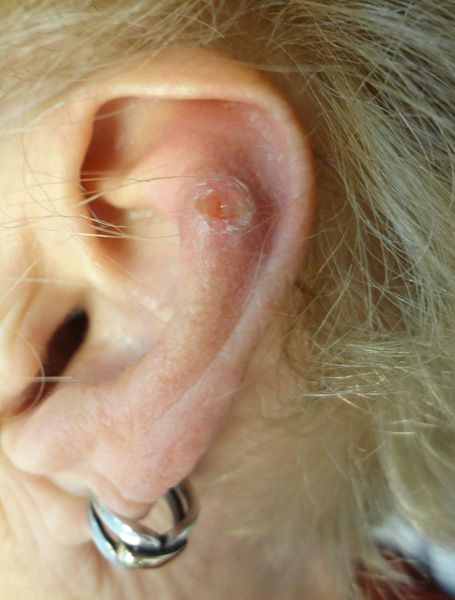
A 71-year-old woman sought medical attention for a chronically painful ear lesion.
Key point: Examination revealed a 0.5-cm ulcerated and tender nodule on the superior pole of the antihelix of the left ear. This strongly suggests basal cell or squamous cell carcinoma. A biopsy, however, disclosed only an intense inflammatory infiltrate extending to and into the cartilage. This is characteristic for chondrodermatitis nodularis helicis (CNH)-an idiopathic inflammatory disorder of the external ear.
Treatment: Surgical debridement, coupled with injection of dilute triamcinolone acetonide suspension (2 to 3 mg/mL) may be employed. In this case, intralesional corticosteroid injections to the ulcer base and lesion periphery were performed after the biopsy. The area healed and did not recur.
Note: CNH is usually painful, whereas skin cancer of the pinna is rarely painful.


