- Clinical Technology
- Adult Immunization
- Hepatology
- Pediatric Immunization
- Screening
- Psychiatry
- Allergy
- Women's Health
- Cardiology
- Pediatrics
- Dermatology
- Endocrinology
- Pain Management
- Gastroenterology
- Infectious Disease
- Obesity Medicine
- Rheumatology
- Nephrology
- Neurology
- Pulmonology
Catscratch Disease Presenting as Acute Respiratory Distress
Superficial adenopathy is the most common symptom ofcatscratch disease (CSD) attributed to Bartonella henselaeinfection. More complicated adenopathy with pulmonaryinvolvement can occur. We report a case of a 15-year-oldboy with pleural symptoms related to B henselae–associatedCSD. [Infect Med. 2008;25:248-250]
Bartonella henselae is the bacterium that causes catscratch disease (CSD) and is the most common species of Bartonella seen in immunocompetent patients with the infection. Although superficial adenopathy is the most common symptom, a range of other symptoms, including pulmonary involvement, can occur. Painful axial adenopathy was the presenting symptom in the patient in the case we describe here. Examination of pleural fluid and radiographs suggested inflammatory processes within the lungs.
Case report
A 15-year-old boy was admitted to the department of infectious diseases for respiratory distress. He had no history of respiratory distress but had had allergic rhinitis with untreated effort-related asthma. Two weeks before the first clinical symptoms appeared, the patient was wounded on the dorsal surface of his right arm by a pet cat. One week later, progressive dyspnea developed, which resulted in the patient's admission to the ICU for acute respiratory distress.
During the physical examination, the patient's respiration rate was more than 40 breaths per minute. The only lung abnormality noted was the presence of painful adenopathy in the right axillary area. The lesions were mobile, fluctuant, and more than 3 cm in diameter. The few biological abnormalities that were seen included a white blood cell count that was slightly higher than normal (10,192/& mu;L), a high neutrophil count (1900/& mu;L), and a high C-reactive protein level (38 mg/L). Blood gas examination showed hypoxemia (PO2, 50 mm Hg) and hypocapnia (PCO2, 29 mm Hg). Results of serological tests were negative for Toxoplasma gondii, Epstein-Barrvirus, Chlamydia pneumoniae, Mycoplasma pneumoniae, and Legionnella pneumophila.
Findings from bronchoalveolar lavage performed on admission were normal. Culture of a pleural fluid specimen obtained during the second week of hospitalization remained sterile, but it was performed after the patient had started antibiotic treatment. The pleural fluid exhibited an inflammatory profile, but findings from cerebrospinal fluid examination were normal.
The right axillary adenopathy was aspirated. Cytological examination showed a nonspecific inflammatory profile. Results of a Gram stain were normal. Cultures of node aspirates and alveolar, pleural, and cerebrospinal fluid were negative for aerobic and anaerobic microorganisms.
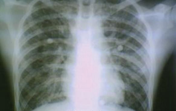
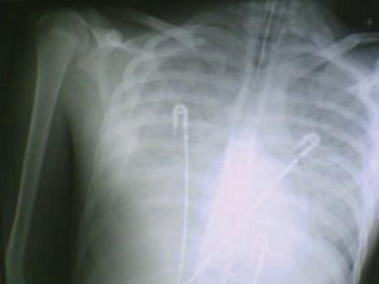
Chest radiographs, obtained daily, showed diffuse bilateral alveolointerstitial infiltrates (Figure 1); a moderate bilateral pleural effusion occurred during the second week of hospitalization (Figure 2).

Figure 1 - This radiograph illustrates diffuse bilateral alveolo-interstitial infiltrates.

Figure 2 - The extensive lesions shown here are responsible for the patient's acute respiratorydistress syndrome. Pleural effusion is most prominent on the right side of the chest.
Immunofluorescence assay was positive for B henselae (IgG, 1/50; IgM, 1/50). Results of the polymerase chain reaction (PCR) assay performed on the fluid that was aspirated from the axillary adenopathy also were positive for B henselae. Therefore, the definite diagnosis was acute respiratory distress syndrome associated with B henselae CSD.
The patient was initially treated with a combination of intravenous amoxicillin and clarithromycin. Two days after the initial examination, the patient experienced an epileptic subintrant crisis that lasted a few minutes and required intubation with mechanical ventilatory assistance. The antibiotic regimen wasswitched to intravenous amoxicillin/ clavulanate and linezolid to provide a broader spectrum of activity (specifically, activity against methicillin- resistant staphylococci and gram-negative bacilli).
The patient achieved a full recovery without complications or adverse effects after 7 days of intravenous therapy. However, adenophlegmon with spontaneous effusion developed in the right axillary area 1 month later. Local treatment was given, and the condition resolved without further complication.
Discussion
Bartonella is a gram-negative facultative intracellular bacillus; the genus includes 13 distinct species. The clinical symptoms of CSD appear 1 to 7 weeks after the inoculation of the skin lesion, usually caused by a cat scratch. The clinical signs are frequently limited to superficial adenopathy (50% to 70% of cases). In more than 10% of cases, numerous related syndromes can be observed, including Parinaud oculoglandular syndrome, cerebral abscess, polyneuropathy, retinal neuropathy, peripheral thrombopathy, erythema nodosum, erythema migrans, urethritis, splenic and hepatic granuloma, osteomyelitis, pulmonary and pleural effusion, endocarditis, and bacteremia.1 Encephalopathy has been reported to occur in about 2% of cases.1
In most cases, the diagnosis of CSD is based on serological tests.2 PCR assay is useful in identifying atypical forms.1,3 Recent studies have established that the serological sensitivity of indirect fluorescence assay is about 50%.2,4 Cross-reactions between different species of Bartonella, Chlamydia, and Coxiella may reduce the specificity of the serological test results.3 PCR techniques seem to have higher sensitivity and specificity than serological techniques such as enzyme-linked immunosorbent assay.1,4-6
Antibiotics that have been used in the treatment of CSD include tetracyclines, macrolides, rifampicin, fluoroquinolones, trimethoprim/sulfamethoxazole, aminoglycosides, and phenicoles.7 The role of antibiotictreatment, however, is controversial. 1 The clinical outcome is usually favorable for patients with CSDassociated pulmonary abnormalities, but the effectiveness of antibiotic therapy is not clear.8 CSD can resolve without antibiotic therapy. Although B henselae is susceptible to many antibiotics in vivo, the clinical signs and symptoms of CSD do not always resolve quickly. According to some authors, pulmonary involvement in immunocompetent patients may be attributed to lesions of the vascular endothelium secondary to bacterial infection.8 Other authors contend that the antigenic stimulation may result in clinical manifestations in the immunocompetent host.1,9,10
Although pulmonary involvement in CSD is uncommon, CSD should be considered in the differential diagnosis in immunocompetent patients who present with acute respiratory distress syndrome and who have a history of contact with cats. PCR is a reliable and rapid method for confirming the diagnosis.
References:
- Regnery R, Tappero J. Unraveling mysteries associated with cat-scratch disease, bacillary angiomatosis, and related syndromes. Emerg Infect Dis. 1995;1:16-21.
- Regnery R, Olson J, Perkins B, Bibb W. Serological response to "Rochalimaea henselae" antigen in suspected cat-scratch disease. Lancet. 1992; 339:1443-1445.
- Dupon M, Savin De Larclause AM, Brouqui P, et al. Evaluation of serological response to Bartonella henselae, Bartonella quintana and Afipia felis antigens in 64 patients with suspected catscratch disease. Scand J Infect Dis. 1996;28:361- 366
- Bergmans A, Peeters M, Schellekens J, et al. Pitfalls and fallacies of cat scratch disease serology: evaluation of Bartonella henselae-based indirect fluorescence assay and enzyme linked immunoassay. J Clin Microbiol. 1997;35:1931-1937.
- Hansmann Y, DeMartino S, Piémont Y, et al. Diagnosis of cat scratch disease with detection of Bartonella henselae by PCR: a study of patients with lymph node enlargement. J Clin Microbiol. 2005;43:3800-3806.
- Anderson B, Sims K, Regnery R, et al. Detection of Rochalimaea henselaeDNAin specimens from cat scratch disease patients by PCR. J Clin Microbiol. 1994;32:942-948.
- Rolain JM, Brouqui P, Koehler JE, et al. Recommendations for treatment of human infections caused by Bartonella species. Antimicrob Agents Chemother. 2004;48:1921-1933.
- Abbasi S, Chesney PJ. Pulmonary manifestations of cat-scratch disease: a case report and review of the literature. Pediatr Infect Dis J. 1995; 14:547-548.
- Margileth AM. Recent advances in diagnosis and treatment of cat scratch disease. Curr Infect Dis Rep. 2000;2:141-146.
- Margileth AM, Baehren DF. Chest-wall abscess due to cat scratch disease (CSD) in an adult with antibodies to Bartonella clarridgeiae: case report and review of the thoracopulmonary manifestations of CSD. Clin Infect Dis. 1998;27:353- 357.
