- Clinical Technology
- Adult Immunization
- Hepatology
- Pediatric Immunization
- Screening
- Psychiatry
- Allergy
- Women's Health
- Cardiology
- Pediatrics
- Dermatology
- Endocrinology
- Pain Management
- Gastroenterology
- Infectious Disease
- Obesity Medicine
- Rheumatology
- Nephrology
- Neurology
- Pulmonology
A Case of Giant Colonic Diverticulum

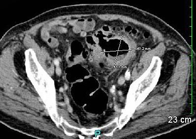
An 88-year-old man with a history of hypertension, coronary artery disease, atrial fibrillation, and hypothyroidism presented to his primary care physician complaining of malaise and a weight loss of 20 lbs over the past few months. His physical examination was unremarkable. Serum chemistries, however, revealed anemia-hematocrit, 28% (normal range, 41% - 51%)]. Abdominal CT revealed a 5- to 6-cm lesion in the pelvis suggestive of either a walled-off abscess or a diverticulum (Figure). After a surgical consultation, the patient was admitted to the hospital for further evaluation.
A colonoscopy found a sigmoid stricture and diverticulosis within the visualized portion of the sigmoid colon. Broad-spectrum antibiotics were initiated and a sigmoid colectomy was performed the following day. Surgical findings included a large sigmoid diverticulum surrounded by severe inflammation. Histologic analysis revealed a pseudocyst consisting of inflammatory tissue surrounding the sigmoid colon with a communication to the anti-mesenteric surface of the colon. The rectum was closed with a Hartmann pouch and an end-colostomy was placed. A diagnosis was made of giant colonic diverticulum (4 x 3 x 2.5 cm). After several weeks of rehabilitation at a skilled nursing facility, the patient was discharged. Our patient is currently 91 years of age, living at home, and his stoma is managed by a caregiver. He does have a moderate peristomal hernia but has declined surgical closure.
Discussion
Giant colonic diverticulum (GCD) is a rare and extreme manifestation on the continuum of diverticular disease. It was described in the early 1950s as a “solitary air cyst.”1 Presentation can be acute, however, symptomatology most often follows an indolent course. Classification is based on size and histologic findings. GCDs are defined as lesions 4 cm or larger in diameter.2 There are 2 histologic types. A type I GCD, as in this case, also called a pseudo diverticulum, is most likely associated with conventional diverticular disease and occurs when a contained perforation forms an air-filled pseudocapsule that continues to enlarge. Approximately 87% of GCDs are classified as Type I.3 A type II, or true diverticulum, contains all layers of bowel wall and structurally resembles a duplication cyst.4 True GCDs are rare and are thought to be congenital.
Nearly all (~90%) of GCDs, regardless of type, are found within the sigmoid colon; 25% communicate with the lumen; and the majority occur on the anti-mesenteric side of the colon.5,6 Most GCDs measure between 7 and 15 cm. As with diverticular disease, this usually affects patients in their 60’s and 70’s.7 Up to 70% of GCDs are palpable on examination.7 These patients can present acutely, but more often onset is insidious with gradual development of chronic pain.6
GCDs are detectable on abdominal x-ray and appear as large air-filled structures (the “balloon sign”). Diagnosis is most often based on CT findings, and CT imaging allows better visualization of surrounding structures.7,8 CT with water soluble contrast may help detect communication with the colon. Colonoscopy may provide visualization of other lesions but adds little additional information about GCD.3
Initial management includes IV hydration, antibiotics, and analgesics. Unless contraindicated by medical comorbidities, surgical resection with or without primary anastamosis is indicated.6,9 Selected cases of true diverticula may be adequately removed by diverticulectomy alone.6,9
A palpable abdominal mass is usually an ominous finding. GCD, however, is a potentially curable condition that tends to occur in members of the age group often given a diagnosis of a malignancy. Therefore, aggressive work-up and early surgical intervention are warranted.
References:
References
1. Hughes WL, Greene RC: Solitary air cyst of peritoneal cavity. Arch Surg. 1953;74:931-936.
2. Nicholson Y, Angel LP, Caliendo, FC, Procaccino, JA. Giant colonic diverticula. Surgical Rounds. 2007:224-228.
3. Mehta, DC, Baum, JA, Dave, PB, Gumaste, VV. Giant sigmoid diverticulum: a report of two cases and endoscopic recognition. Am J Gastroenterol 1996;91:1269-1271.
4. Ryckman, FC, Glenn JD, Moazam F. Spontaneous perforation of a colonic duplication. Dis Colon Rectum. 1983;26:287-289.
5. Sagar, S. Giant solitary diverticulum of the transverse colon with diverticulosis. Br J Clin Pract. 1973;27:145-146.
6. Maresca L, Maresca C, Erickson E. Giant sigmoid diverticulum: a report of a case. Dis Colon Rectum. 1981;24:191-195.
7. Chaiyasate K, Yavuzer R, Mittal V. Giant sigmoid diverticulum. Surgery. 2006;139(2):276-277.
8. Rosenberg RF, Naidich JB. Plain film recognition of giant colonic diverticulum. Am J Gastroentero.l 1981;76:59-69.9. Al-Jurf AS, Foucar E. Uncommon features of giant colonic diverticula. Dis Colon Rectum. 1983;26:808-813.
